
Leveraging artificial intelligence and machine learning for unraveling pathogenesis and advancing precision medicine in autoimmune diseases
-
GRAPHICAL ABSTRACT

 DownLoad:
Full size image
DownLoad:
Full size image
-
PUBLIC SUMMARY
-
Artificial intelligence (AI) and machine learning (ML) accelerate understanding of the immune system.
AI and ML provide new perspectives for diagnosing and treating autoimmune diseases.
AI and ML assist in predicting treatment responses and contribute to drug development.
-
-
ABSTRACT
Autoimmune diseases (AIDs) are intricate disorders in which the immune system mistakenly attacks the body’s own tissues. Recent advancements in omics technologies, as well as artificial intelligence (AI) and machine learning (ML), have significantly deepened our understanding of AIDs. AI, which mimics intelligent behavior to perform complex tasks, is transforming diagnostic approaches, risk assessments, and health management strategies. High-throughput technologies, including microarrays and single-cell RNA sequencing (scRNA-seq), now allow researchers to assess gene expression profiles, offering valuable insights into disease mechanisms. When combined, AI and ML facilitate the integration of multimodal omics data, aiding in the identification of key regulatory networks, disease subtypes, and potential biomarkers. In basic research, ML investigates immune cell functions, B cell receptor (BCR) and T cell receptor (TCR) interactions, and the major histocompatibility complex (MHC). Clinically, AI supports diagnosis, treatment response prediction, and outcome forecasting. It enables precise patient stratification in major AIDs, such as rheumatoid arthritis (RA), systemic lupus erythematosus (SLE), and systemic sclerosis (SSc), through the integration of clinical, imaging, and multi-omics data. In drug development, AI is revolutionizing traditional research models by assisting in the design of small molecules, engineering antibodies, and developing innovative therapies. However, challenges regarding data quality, model interpretability, and ethical considerations persist. Despite these hurdles, the integration of AI and ML is anticipated to propel advances in precision medicine for AIDs. This review highlights the latest applications of AI and ML in AIDs, focusing on disease mechanisms, diagnostics, treatment prediction, and drug development. -

-
REFERENCES
[1] Smolen J. S., Aletaha D. and McInnes I. B. (2016). Rheumatoid arthritis. Lancet 388:2023−2038. DOI:10.1016/s0140-6736(16)30173-8 [2] Wang L., Wang F. S. and Gershwin M. E. (2015). Human autoimmune diseases: a comprehensive update. J. Intern. Med. 278:369−395. DOI:10.1111/joim.12395 [3] Sanz I. and Lund F. (2019). Complexity and heterogeneity - the defining features of autoimmune disease. Curr. Opin. Immunol. 61:iii−vi. DOI:10.1016/j.coi.2019.11.006 [4] Davidson A. and Diamond B. (2001). Autoimmune diseases. N. Engl. J. Med. 345:340−350. DOI:10.1056/nejm200108023450506 [5] Baker K. F. and Isaacs J. D. (2018). Novel therapies for immune-mediated inflammatory diseases: What can we learn from their use in rheumatoid arthritis, spondyloarthritis, systemic lupus erythematosus, psoriasis, Crohn's disease and ulcerative colitis. Ann. Rheum. Dis. 77:175−187. DOI:10.1136/annrheumdis-2017-211555 [6] Fugger L., Jensen L. T. and Rossjohn J. (2020). Challenges, Progress, and Prospects of Developing Therapies to Treat Autoimmune Diseases. Cell 181:63−80. DOI:10.1016/j.cell.2020.03.007 [7] Anchang C. G., Xu C., Raimondo M. G., et al. (2021). The Potential of OMICs Technologies for the Treatment of Immune-Mediated Inflammatory Diseases. Int. J. Mol. Sci. 22. DOI:10.3390/ijms22147506 [8] Chu X., Zhang B., Koeken V., et al. (2021). Multi-Omics Approaches in Immunological Research. Front. Immunol. 12:668045. DOI:10.3389/fimmu.2021.668045 [9] Nagaoka K., Shirai M., Taniguchi K., et al. (2020). Deep immunophenotyping at the single-cell level identifies a combination of anti-IL-17 and checkpoint blockade as an effective treatment in a preclinical model of data-guided personalized immunotherapy. J. Immunother. Cancer 8. DOI:10.1136/jitc-2020-001358 [10] Buniello A., MacArthur J. A. L., Cerezo M., et al. (2019). The NHGRI-EBI GWAS Catalog of published genome-wide association studies, targeted arrays and summary statistics 2019. Nucleic Acids Res. 47:D1005−d1012. DOI:10.1093/nar/gky1120 [11] Sewell G. W. and Kaser A. (2022). Interleukin-23 in the pathogenesis of inflammatory bowel disease and implications for therapeutic intervention. J. Crohns Colitis 16:ii3−ii19. DOI:10.1093/ecco-jcc/jjac034 [12] Yin X., Bose D., Kwon A., et al. (2022). Integrating transcriptomics, metabolomics, and GWAS helps reveal molecular mechanisms for metabolite levels and disease risk. Am. J. Hum. Genet. 109:1727−1741. DOI:10.1016/j.ajhg.2022.08.007 [13] Mountjoy E., Schmidt E. M., Carmona M., et al. (2021). An open approach to systematically prioritize causal variants and genes at all published human GWAS trait-associated loci. Nat. Genet. 53:1527−1533. DOI:10.1038/s41588-021-00945-5 [14] Na F., Pan X., Chen J., et al. (2022). KMT2C deficiency promotes small cell lung cancer metastasis through DNMT3A-mediated epigenetic reprogramming. Nat. Cancer 3:753−767. DOI:10.1038/s43018-022-00361-6 [15] Abell N. S., DeGorter M. K., Gloudemans M. J., et al. (2022). Multiple causal variants underlie genetic associations in humans. Science 375:1247−1254. DOI:10.1126/science.abj5117 [16] Assum I., Krause J., Scheinhardt M. O., et al. (2022). Tissue-specific multi-omics analysis of atrial fibrillation. Nat. Commun. 13:441. DOI:10.1038/s41467-022-27953-1 [17] Suhre K., Arnold M., Bhagwat A. M., et al. (2017). Erratum: Connecting genetic risk to disease end points through the human blood plasma proteome. Nat. Commun. 8:15345. DOI:10.1038/ncomms15345 [18] Gulfidan G., Soylu M., Demirel D., et al. (2022). Systems biomarkers for papillary thyroid cancer prognosis and treatment through multi-omics networks. Arch. Biochem. Biophys. 715:109085. DOI:10.1016/j.abb.2021.109085 [19] Lu J., Zhang Y., Sun M., et al. (2021). Multi-omics analysis of fatty acid metabolism in thyroid carcinoma. Front. Oncol. 11:737127. DOI:10.3389/fonc.2021.737127 [20] Raivola J., Dini A., Karvonen H., et al. (2022). Multiomics characterization implicates PTK7 in ovarian cancer EMT and cell plasticity and offers strategies for therapeutic intervention. Cell Death Dis. 13:714. DOI:10.1038/s41419-022-05161-5 [21] Martorell-Marugán J., López-Domínguez R., García-Moreno A., et al. (2021). A comprehensive database for integrated analysis of omics data in autoimmune diseases. BMC Bioinformatics 22:343. DOI:10.1186/s12859-021-04268-4 [22] Rein P. and Mueller R. B. (2017). Treatment with biologicals in rheumatoid arthritis: An overview. Rheumatol. Ther. 4:247−261. DOI:10.1007/s40744-017-0073-3 [23] Burmester G. R. and Pope J. E. (2017). Novel treatment strategies in rheumatoid arthritis. Lancet 389:2338−2348. DOI:10.1016/s0140-6736(17)31491-5 [24] Deo R. C. (2015). Machine learning in medicine. Circulation 132:1920−1930. DOI:10.1161/circulationaha.115.001593 [25] LeCun Y., Bengio Y. and Hinton G. (2015). Deep learning. Nature 521:436−444. DOI:10.1038/nature14539 [26] Liu F., Jang H., Kijowski R., et al. (2018). Deep learning MR imaging-based attenuation correction for PET/MR imaging. Radiology 286:676−684. DOI:10.1148/radiol.2017170700 [27] Dey D., Slomka P. J., Leeson P., et al. (2019). Artificial intelligence in cardiovascular imaging: JACC state-of-the-art review. J. Am. Coll. Cardiol. 73:1317−1335. DOI:10.1016/j.jacc.2018.12.054 [28] Su X., Chen N., Sun H., et al. (2020). Automated machine learning based on radiomics features predicts H3 K27M mutation in midline gliomas of the brain. Neuro. Oncol. 22:393−401. DOI:10.1093/neuonc/noz184 [29] Aerts H. J. (2016). The potential of radiomic-based phenotyping in precision medicine: A review. JAMA Oncol. 2:1636−1642. DOI:10.1001/jamaoncol.2016.2631 [30] Fiocchi C. and Iliopoulos D. (2020). What's new in IBD therapy: An "omics network" approach. Pharmacol. Res. 159:104886. DOI:10.1016/j.phrs.2020.104886 [31] Hosny A., Parmar C., Quackenbush J., et al. (2018). Artificial intelligence in radiology. Nat. Rev. Cancer 18:500−510. DOI:10.1038/s41568-018-0016-5 [32] Notin P., Rollins N., Gal Y., et al. (2024). Machine learning for functional protein design. Nat. Biotechnol. 42:216−228. DOI:10.1038/s41587-024-02127-0 [33] Badillo S., Banfai B., Birzele F., et al. (2020). An introduction to machine learning. Clin. Pharmacol. Ther. 107:871−885. DOI:10.1002/cpt.1796 [34] Choi R. Y., Coyner A. S., Kalpathy-Cramer J., et al. (2020). Introduction to machine learning, neural networks, and deep learning. Transl. Vis. Sci. Technol. 9:14. DOI:10.1167/tvst.9.2.14 [35] Min S., Lee B. and Yoon S. (2017). Deep learning in bioinformatics. Brief. Bioinform. 18:851−869. DOI:10.1093/bib/bbw068 [36] Domingos P. (2012). A few useful things to know about machine learning. Communications of the ACM 55:78−87. DOI:10.1145/2347736.2347755 [37] Libbrecht M. W. and Noble W. S. (2015). Machine learning applications in genetics and genomics. Nat. Rev. Genet. 16:321−332. DOI:10.1038/nrg3920 [38] Brown M. P., Grundy W. N., Lin D., et al. (2000). Knowledge-based analysis of microarray gene expression data by using support vector machines. Proc. Natl. Acad. Sci. U. S. A. 97:262−267. DOI:10.1073/pnas.97.1.262 [39] Shah S. P., McVicker G. P., Mackworth A. K., et al. (2003). GeneComber: Combining outputs of gene prediction programs for improved results. Bioinformatics 19:1296−1297. DOI:10.1093/bioinformatics/btg139 [40] Lewis B. P., Shih I. H., Jones-Rhoades M. W., et al. (2003). Prediction of mammalian microRNA targets. Cell 115:787−798. DOI:10.1016/s0092-8674(03)01018-3 [41] Karwasra R., Sharma S., Sharma I., et al. (2024). Autoimmune autonomic disorder: AI-based diagnosis and prognosis. In Artificial Intelligence and Autoimmune Diseases: Applications in the Diagnosis, Prognosis, and Therapeutics (Springer), pp. 77-98. [42] Lezcano-Valverde J. M., Salazar F., León L., et al. (2017). Development and validation of a multivariate predictive model for rheumatoid arthritis mortality using a machine learning approach. Sci. Rep. 7:10189. DOI:10.1038/s41598-017-10558-w [43] Guan Y., Zhang H., Quang D., et al. (2019). Machine learning to predict anti-tumor necrosis factor drug responses of rheumatoid arthritis patients by integrating clinical and genetic markers. Arthritis Rheumatol. 71:1987−1996. DOI:10.1002/art.41056 [44] Ching T., Himmelstein D. S., Beaulieu-Jones B. K., et al. (2018). Opportunities and obstacles for deep learning in biology and medicine. J. R. Soc. Interface 15:20170387. DOI:10.1098/rsif.2017.0387 [45] Schlereth M., Mutlu M. Y., Utz J., et al. (2024). Deep learning-based classification of erosion, synovitis and osteitis in hand MRI of patients with inflammatory arthritis. RMD Open 10:e004273. DOI:10.1136/rmdopen-2024-004273 [46] Zou J., Huss M., Abid A., et al. (2019). A primer on deep learning in genomics. Nat. Genet. 51:12−18. DOI:10.1038/s41588-018-0295-5 [47] Eraslan G., Avsec Ž., Gagneur J., et al. (2019). Deep learning: New computational modelling techniques for genomics. Nat. Rev. Genet. 20:389−403. DOI:10.1038/s41576-019-0122-6 [48] Ringnér M. (2008). What is principal component analysis. Nat. Biotechnol. 26:303−304. DOI:10.1038/nbt0308-303 [49] Kobak D. and Berens P. (2019). The art of using t-SNE for single-cell transcriptomics. Nat. Commun. 10:5416. DOI:10.1038/s41467-019-13056-x [50] Langfelder P. and Horvath S. (2008). WGCNA: An R package for weighted correlation network analysis. BMC Bioinformatics 9:559. DOI:10.1186/1471-2105-9-559 [51] Satija R., Farrell J. A., Gennert D., et al. (2015). Spatial reconstruction of single-cell gene expression data. Nat. Biotechnol. 33:495−502. DOI:10.1038/nbt.3192 [52] Bader G. D. and Hogue C. W. (2003). An automated method for finding molecular complexes in large protein interaction networks. BMC Bioinformatics 4:2. DOI:10.1186/1471-2105-4-2 [53] Margolin A. A., Nemenman I., Basso K., et al. (2006). ARACNE: an algorithm for the reconstruction of gene regulatory networks in a mammalian cellular context. BMC Bioinformatics 7 Suppl 1:S7. DOI:10.1186/1471-2105-7-s1-s7 [54] Wang W., Yang S., Zhang X., et al. (2014). Drug repositioning by integrating target information through a heterogeneous network model. Bioinformatics 30:2923−2930. DOI:10.1093/bioinformatics/btu403 [55] Warde-Farley D., Donaldson S. L., Comes O., et al. (2010). The GeneMANIA prediction server: biological network integration for gene prioritization and predicting gene function. Nucleic Acids Res. 38:W214−220. DOI:10.1093/nar/gkq537 [56] Banchereau R., Hong S., Cantarel B., et al. (2016). Personalized immunomonitoring uncovers molecular networks that stratify lupus patients. Cell 165:551−565. DOI:10.1016/j.cell.2016.03.008 [57] Tasaki S., Suzuki K., Kassai Y., et al. (2018). Multi-omics monitoring of drug response in rheumatoid arthritis in pursuit of molecular remission. Nat. Commun. 9:2755. DOI:10.1038/s41467-018-05044-4 [58] Zhang F., Wei K., Slowikowski K., et al. (2019). Defining inflammatory cell states in rheumatoid arthritis joint synovial tissues by integrating single-cell transcriptomics and mass cytometry. Nat. Immunol. 20:928−942. DOI:10.1038/s41590-019-0378-1 [59] Solomon D. H., Guan H., Johansson F. D., et al. (2023). Assessing clusters of comorbidities in rheumatoid arthritis: A machine learning approach. Arthritis Res. Ther. 25:224. DOI:10.1186/s13075-023-03191-8 [60] Lipton Z. C., Berkowitz J. and Elkan C. (2015). A critical review of recurrent neural networks for sequence learning. arXiv Preprint arXiv:1506.00019. DOI:10.48550/arXiv.1506.00019 [61] Graves A. (2012). Long Short-Term Memory. In Supervised Sequence Labelling with Recurrent Neural Networks (Springer), pp:37-45. DOI:10.1007/978-3-642-24797-2_4 [62] Vincent P., Larochelle H., Lajoie I., et al. (2010). Stacked denoising autoencoders: Learning useful representations in a deep network with a local denoising criterion. J. Mach. Learn. Res. 11:3371−3408. DOI:10.5555/1756006.1953039 [63] Goodfellow I., Pouget-Abadie J., Mirza M., et al. (2014). Generative adversarial nets. Advances in neural information processing systems 27. https://proceedings.neurips.cc/paper_files/paper/2014/file/f033ed80deb0234979a61f95710dbe25-Paper.pdf [64] Kingma D. P. and Welling M. (2013). Auto-encoding variational bayes. arXiv preprint arXiv:1312.6114. DOI:10.48550/arXiv.1312.6114 [65] Vaswani A., Shazeer N., Parmar N., et al. (2017). Attention is all you need. arXiv preprint arXiv:1706.03762. DOI:10.48550/arXiv.1706.03762 [66] Jumper J., Evans R., Pritzel A., et al. (2021). Highly accurate protein structure prediction with AlphaFold. Nature 596:583−589. DOI:10.1038/s41586-021-03819-2 [67] Abramson J., Adler J., Dunger J., et al. (2024). Accurate structure prediction of biomolecular interactions with AlphaFold 3. Nature 630:493−500. DOI:10.1038/s41586-024-07487-w [68] Ji Y., Zhou Z., Liu H., et al. (2021). DNABERT: Pre-trained bidirectional encoder representations from transformers model for DNA-language in genome. Bioinformatics 37:2112−2120. DOI:10.1093/bioinformatics/btab083 [69] Huang K., Xiao C., Glass L. M., et al. (2021). MolTrans: Molecular interaction transformer for drug-target interaction prediction. Bioinformatics 37:830−836. DOI:10.1093/bioinformatics/btaa880 [70] Elnaggar A., Heinzinger M., Dallago C., et al. (2022). ProtTrans: Toward understanding the language of life through self-supervised learning. IEEE Trans. Pattern Anal. Mach. Intell. 44:7112−7127. DOI:10.1109/tpami.2021.3095381 [71] Zheng Y. and Gao G. F. (2023). Geneformer: A deep learning model for exploring gene networks. Sci. China Life Sci. 66:2952−2954. DOI:10.1007/s11427-023-2431-x [72] Wu X., Shuai W., Chen C., et al. (2023). Rapid screening for autoimmune diseases using Fourier transform infrared spectroscopy and deep learning algorithms. Front. Immunol. 14:1328228. DOI:10.3389/fimmu.2023.1328228 [73] Subramani J., Kumar G. S. and Gadekallu T. R. (2024). Gene-based predictive modelling for enhanced detection of systemic lupus erythematosus using CNN-based DL algorithm. Diagnostics (Basel) 14:1339. DOI:10.3390/diagnostics14131339 [74] Huang C., Chen W., Liu B., et al. (2022). Transformer-based deep-learning algorithm for discriminating demyelinating diseases of the central nervous system with neuroimaging. Front. Immunol. 13:897959. DOI:10.3389/fimmu.2022.897959 [75] Capurro N., Pastore V. P., Touijer L., et al. (2024). A deep learning approach to direct immunofluorescence pattern recognition in autoimmune bullous diseases. Br. J. Dermatol. 191:261−266. DOI:10.1093/bjd/ljae142 [76] Li Q., Yang Z., Chen K., et al. (2024). Human-multimodal deep learning collaboration in 'precise' diagnosis of lupus erythematosus subtypes and similar skin diseases. J. Eur. Acad. Dermatol. Venereol. 38:2268−2279. DOI:10.1111/jdv.20031 [77] Ercan C., Kordy K., Knuuttila A., et al. (2024). A deep-learning-based model for assessment of autoimmune hepatitis from histology: AI(H). Virchows Arch. 485:1095−1105. DOI:10.1007/s00428-024-03841-5 [78] Rabhi S., Blanchard F., Diallo A. M., et al. (2022). Temporal deep learning framework for retinopathy prediction in patients with type 1 diabetes. Artif. Intell. Med. 133:102408. DOI:10.1016/j.artmed.2022.102408 [79] Dietterich T. G. (2000). Ensemble Methods in Machine Learning. In Multiple Classifier Systems (Springer), pp:1-15. DOI:10.1007/3-540-45014-9_1 [80] Wu Z., Ramsundar B., Feinberg E. N., et al. (2018). MoleculeNet: A benchmark for molecular machine learning. Chem. Sci. 9:513−530. DOI:10.1039/c7sc02664a [81] Lin Y., Cao Y., Kim H. J., et al. (2020). scClassify: Sample size estimation and multiscale classification of cells using single and multiple reference. Mol. Syst. Biol. 16:e9389. DOI:10.15252/msb.20199389 [82] Alsentzer E., Murphy J. R., Boag W., et al. (2019). Publicly available clinical BERT embeddings. arXiv preprint arXiv:1904.03323. DOI:10.48550/arXiv.1904.03323 [83] Cui H., Wang C., Maan H., et al. (2024). scGPT: Toward building a foundation model for single-cell multi-omics using generative AI. Nat. Methods 21:1470−1480. DOI:10.1038/s41592-024-02201-0 [84] Luo R., Sun L., Xia Y., et al. (2022). BioGPT: Generative pre-trained transformer for biomedical text generation and mining. Brief. Bioinform. 23:bbac409. DOI:10.1093/bib/bbac409 [85] Pun F. W., Ozerov I. V. and Zhavoronkov A. (2023). AI-powered therapeutic target discovery. Trends Pharmacol. Sci. 44:561−572. DOI:10.1016/j.tips.2023.06.010 [86] Ahmed E., Heinzinger M., Dallago C., et al. (2020). Prottrans: Towards cracking the language of life’s code through self-supervised deep learning and high performance computing. arXiv preprint arXiv:2007.06225. DOI:10.48550/arXiv.2007.06225 [87] Xu J., Glicksberg B. S., Su C., et al. (2021). Federated learning for healthcare informatics. J. Healthc. Inform. Res. 5:1−19. DOI:10.1007/s41666-020-00082-4 [88] Avsec Ž., Agarwal V., Visentin D., et al. (2021). Effective gene expression prediction from sequence by integrating long-range interactions. Nat. Methods 18:1196−1203. DOI:10.1038/s41592-021-01252-x [89] Bunne C., Roohani Y., Rosen Y., et al. (2024). How to build the virtual cell with artificial intelligence: Priorities and opportunities. Cell 187:7045−7063. DOI:10.1016/j.cell.2024.11.015 [90] Culos A., Tsai A. S., Stanley N., et al. (2020). Integration of mechanistic immunological knowledge into a machine learning pipeline improves predictions. Nat. Mach. Intell. 2:619−628. DOI:10.1038/s42256-020-00232-8 [91] Reynisson B., Alvarez B., Paul S., et al. (2020). NetMHCpan-4.1 and NetMHCIIpan-4.0: Improved predictions of MHC antigen presentation by concurrent motif deconvolution and integration of MS MHC eluted ligand data. Nucleic Acids Res. 48:W449-W454. DOI:10.1093/nar/gkaa379 [92] Maslova A., Ramirez R. N., Ma K., et al. (2020). Deep learning of immune cell differentiation. Proc. Natl. Acad. Sci. U. S. A. 117:25655−25666. DOI:10.1073/pnas.2011795117 [93] Evrard M., Becht E., Fonseca R., et al. (2023). Single-cell protein expression profiling resolves circulating and resident memory T cell diversity across tissues and infection contexts. Immunity 56:1664−1680.e1669. DOI:10.1016/j.immuni.2023.06.005 [94] Shaban M., Bai Y., Qiu H., et al. (2024). MAPS: Pathologist-level cell type annotation from tissue images through machine learning. Nat. Commun. 15:28. DOI:10.1038/s41467-023-44188-w [95] Trapnell C., Cacchiarelli D., Grimsby J., et al. (2014). The dynamics and regulators of cell fate decisions are revealed by pseudotemporal ordering of single cells. Nat. Biotechnol. 32:381−386. DOI:10.1038/nbt.2859 [96] Street K., Risso D., Fletcher R. B., et al. (2018). Slingshot: Cell lineage and pseudotime inference for single-cell transcriptomics. BMC Genomics 19:477. DOI:10.1186/s12864-018-4772-0 [97] Setty M., Tadmor M. D., Reich-Zeliger S., et al. (2016). Wishbone identifies bifurcating developmental trajectories from single-cell data. Nat. Biotechnol. 34:637−645. DOI:10.1038/nbt.3569 [98] Chen X., Wang T., Chen L., et al. (2024). Cross-species single-cell analysis uncovers the immunopathological mechanisms associated with IgA nephropathy progression. JCI Insight 9:e173651. DOI:10.1172/jci.insight.173651 [99] Shi Z., Qin H. and Wu H. (2024). Single-cell transcriptome reveals the heterogeneity of T cells in mice with systemic lupus erythematosus and neuronal inflammation. J. Inflamm. Res. 17:11375−11402. DOI:10.2147/jir.S474211 [100] Hu Z., Zheng M., Guo Z., et al. (2024). Single-cell sequencing reveals distinct immune cell features in cutaneous lesions of pemphigus vulgaris and bullous pemphigoid. Clin. Immunol. 263:110219. DOI:10.1016/j.clim.2024.110219 [101] Achar S. R., Bourassa F. X. P., Rademaker T. J., et al. (2022). Universal antigen encoding of T cell activation from high-dimensional cytokine dynamics. Science 376:880−884. DOI:10.1126/science.abl5311 [102] Fu Z. H., He S. Z., Wu Y., et al. (2023). Design and deep learning of synthetic B-cell-specific promoters. Nucleic Acids Res. 51:11967−11979. DOI:10.1093/nar/gkad930 [103] Alghamdi W., Attique M., Alzahrani E., et al. (2022). LBCEPred: A machine learning model to predict linear B-cell epitopes. Brief. Bioinform. 23:bbac035. DOI:10.1093/bib/bbac035 [104] Janocha K., Ling A., Godson A., et al. (2024). Harnessing preference optimisation in protein LMs for hit maturation in cell therapy. arXiv preprint arXiv:2412.01388. DOI:10.48550/arXiv.2412.01388 [105] Ghosh P., Sinha S., Katkar G. D., et al. (2023). Machine learning identifies signatures of macrophage reactivity and tolerance that predict disease outcomes. EBioMedicine 94:104719. DOI:10.1016/j.ebiom.2023.104719 [106] Bassing C. H., Swat W. and Alt F. W. (2002). The mechanism and regulation of chromosomal V(D)J recombination. Cell 109 Suppl:S45-55. DOI:10.1016/s0092-8674(02)00675-x [107] Russell M. L., Souquette A., Levine D. M., et al. (2022). Combining genotypes and T cell receptor distributions to infer genetic loci determining V(D)J recombination probabilities. Elife 11:e73475. DOI:10.7554/eLife.73475 [108] Jiang X., Wang S., Zhou C., et al. (2020). Comprehensive TCR repertoire analysis of CD4(+) T-cell subsets in rheumatoid arthritis. J. Autoimmun. 109:102432. DOI:10.1016/j.jaut.2020.102432 [109] Sidhom J. W., Larman H. B., Pardoll D. M., et al. (2021). DeepTCR is a deep learning framework for revealing sequence concepts within T-cell repertoires. Nat. Commun. 12:1605. DOI:10.1038/s41467-021-21879-w [110] Sidhom J. W., Oliveira G., Ross-MacDonald P., et al. (2022). Deep learning reveals predictive sequence concepts within immune repertoires to immunotherapy. Sci. Adv. 8:eabq5089. DOI:10.1126/sciadv.abq5089 [111] Davidsen K., Olson B. J., DeWitt W. S., 3rd, et al. (2019). Deep generative models for T cell receptor protein sequences. Elife 8:e46935. DOI:10.7554/eLife.46935 [112] Gielis S., Moris P., Bittremieux W., et al. (2019). Detection of enriched T cell epitope specificity in full T cell receptor sequence repertoires. Front. Immunol. 10:2820. DOI:10.3389/fimmu.2019.02820 [113] Croce G., Bobisse S., Moreno D. L., et al. (2024). Deep learning predictions of TCR-epitope interactions reveal epitope-specific chains in dual alpha T cells. Nat. Commun. 15:3211. DOI:10.1038/s41467-024-47461-8 [114] Montemurro A., Schuster V., Povlsen H. R., et al. (2021). NetTCR-2.0 enables accurate prediction of TCR-peptide binding by using paired TCRα and β sequence data. Commun. Biol. 4:1060. DOI:10.1038/s42003-021-02610-3 [115] Lu T., Zhang Z., Zhu J., et al. (2021). Deep learning-based prediction of the T cell receptor-antigen binding specificity. Nat. Mach. Intell. 3:864−875. DOI:10.1038/s42256-021-00383-2 [116] Fast E., Dhar M. and Chen B. (2023). TAPIR: A T-cell receptor language model for predicting rare and novel targets. bioRxiv preprint bioRxiv:2023.09.12.557285. DOI:10.1101/2023.09.12.557285 [117] Diao K., Wu T., Zhao X., et al. Pep2TCR: Accurate prediction of CD4 T cell receptor binding specificity through transfer learning and ensemble approach. iMetaOmics 1:e43. DOI:10.1002/imo2.43 [118] Zhao Y., He B., Xu F., et al. (2023). DeepAIR: A deep learning framework for effective integration of sequence and 3D structure to enable adaptive immune receptor analysis. Sci. Adv. 9:eabo5128. DOI:10.1126/sciadv.abo5128 [119] Isacchini G., Walczak A. M., Mora T., et al. (2021). Deep generative selection models of T and B cell receptor repertoires with soNNia. Proc. Natl. Acad. Sci. U. S. A. 118:e2023141118. DOI:10.1073/pnas.2023141118 [120] Li G., Iyer B., Prasath V. B. S., et al. (2021). DeepImmuno: Deep learning-empowered prediction and generation of immunogenic peptides for T-cell immunity. Brief. Bioinform. 22:bbab160. DOI:10.1093/bib/bbab160 [121] Jiang Y., Huo M. and Cheng Li S. (2023). TEINet: A deep learning framework for prediction of TCR-epitope binding specificity. Brief. Bioinform. 24:ebbad086. DOI:10.1093/bib/bbad086 [122] Lee C. H., Huh J., Buckley P. R., et al. (2023). A robust deep learning workflow to predict CD8 + T-cell epitopes. Genome. Med. 15:70. DOI:10.1186/s13073-023-01225-z [123] Yin R., Ribeiro-Filho H. V., Lin V., et al. (2023). TCRmodel2: High-resolution modeling of T cell receptor recognition using deep learning. Nucleic Acids Res. 51:W569−W576. DOI:10.1093/nar/gkad356 [124] Jiang F., Guo Y., Ma H., et al. (2024). AlphaEpi: Enhancing B Cell Epitope Prediction with AlphaFold 3. Proceedings of the 15th ACM International Conference on Bioinformatics, Computational Biology and Health Informatics 67:1-8. DOI:10.1145/3698587.3701389 [125] Peng X., Lei Y., Feng P., et al. (2023). Characterizing the interaction conformation between T-cell receptors and epitopes with deep learning. Nat. Mach. Intell. 5:395−407. DOI:10.1038/s42256-023-00634-4 [126] Chen J., Zhao B., Lin S., et al. (2024). TEPCAM: Prediction of T-cell receptor-epitope binding specificity via interpretable deep learning. Protein Sci. 33:e4841. DOI:10.1002/pro.4841 [127] Fernando M. M., Stevens C. R., Walsh E. C., et al. (2008). Defining the role of the MHC in autoimmunity: A review and pooled analysis. PLoS Genet. 4:e1000024. DOI:10.1371/journal.pgen.1000024 [128] Matzaraki V., Kumar V., Wijmenga C., et al. (2017). The MHC locus and genetic susceptibility to autoimmune and infectious diseases. Genome Biol. 18:76. DOI:10.1186/s13059-017-1207-1 [129] Shahbazy M., Ramarathinam S. H., Li C., et al. (2024). MHCpLogics: An interactive machine learning-based tool for unsupervised data visualization and cluster analysis of immunopeptidomes. Brief. Bioinform. 25:bbae087. DOI:10.1093/bib/bbae087 [130] Boegel S., Löwer M., Schäfer M., et al. (2013). HLA typing from RNA-Seq sequence reads. Genome Med. 4:1−12. DOI:10.1186/gm403 [131] Wilhelm M., Zolg D. P., Graber M., et al. (2021). Deep learning boosts sensitivity of mass spectrometry-based immunopeptidomics. Nat. Commun. 12:3346. DOI:10.1038/s41467-021-23713-9 [132] O'Donnell T. J., Rubinsteyn A., Bonsack M., et al. (2018). MHCflurry: Open-source class I MHC binding affinity prediction. Cell Syst. 7:129−132.e124. DOI:10.1016/j.cels.2018.05.014 [133] Jurtz V., Paul S., Andreatta M., et al. (2017). NetMHCpan-4.0: Improved peptide–MHC class I interaction predictions integrating eluted ligand and peptide binding affinity data. J. Immunol. 199:3360-3368. DOI:10.4049/jimmunol.1700893 [134] Mei S., Li F., Leier A., et al. (2020). A comprehensive review and performance evaluation of bioinformatics tools for HLA class I peptide-binding prediction. Brief. Bioinform. 21:1119−1135. DOI:10.1093/bib/bbz051 [135] Fanouriakis A., Tziolos N., Bertsias G., et al. (2021). Update οn the diagnosis and management of systemic lupus erythematosus. Ann. Rheum. Dis. 80:14−25. DOI:10.1136/annrheumdis-2020-218272 [136] Emmi G., Bettiol A., Hatemi G., et al. (2024). Behçet's syndrome. Lancet 403:1093−1108. DOI:10.1016/s0140-6736(23)02629-6 [137] Danieli M. G., Brunetto S., Gammeri L., et al. (2024). Machine learning application in autoimmune diseases: State of art and future prospectives. Autoimmun. Rev. 23:103496. DOI:10.1016/j.autrev.2023.103496 [138] Jang S., Kwon E. J. and Lee J. J. (2022). Rheumatoid arthritis: Pathogenic roles of diverse immune cells. Int. J. Mol. Sci. 23:905. DOI:10.3390/ijms23020905 [139] Zhou S. M., Fernandez-Gutierrez F., Kennedy J., et al. (2016). Defining disease phenotypes in primary care electronic health records by a machine learning approach: A case study in identifying rheumatoid arthritis. PLoS One 11:e0154515. DOI:10.1371/journal.pone.0154515 [140] Chin C. Y., Hsieh S. Y. and Tseng V. S. (2018). eDRAM: Effective early disease risk assessment with matrix factorization on a large-scale medical database: A case study on rheumatoid arthritis. PLoS One 13:e0207579. DOI:10.1371/journal.pone.0207579 [141] Maarseveen T. D., Meinderink T., Reinders M. J. T., et al. (2020). Machine learning electronic health record identification of patients with rheumatoid arthritis: Algorithm pipeline development and validation study. JMIR Med. Inform. 8:e23930. DOI:10.2196/23930 [142] Maarseveen T. D., Maurits M. P., Niemantsverdriet E., et al. (2021). Handwork vs machine: A comparison of rheumatoid arthritis patient populations as identified from EHR free-text by diagnosis extraction through machine-learning or traditional criteria-based chart review. Arthritis Res. Ther. 23:174. DOI:10.1186/s13075-021-02553-4 [143] De Stefano L., D'Onofrio B., Gandolfo S., et al. (2023). Seronegative rheumatoid arthritis: One year in review 2023. Clin. Exp. Rheumatol. 41:554−564. DOI:10.55563/clinexprheumatol/go7g26 [144] Scherer H. U., Häupl T. and Burmester G. R. (2020). The etiology of rheumatoid arthritis. J. Autoimmun. 110:102400. DOI:10.1016/j.jaut.2019.102400 [145] Hu C., Dai Z., Xu J., et al. (2022). Proteome profiling identifies serum biomarkers in rheumatoid arthritis. Front. Immunol. 13:865425. DOI:10.3389/fimmu.2022.865425 [146] Yeo L., Adlard N., Biehl M., et al. (2016). Expression of chemokines CXCL4 and CXCL7 by synovial macrophages defines an early stage of rheumatoid arthritis. Ann. Rheum. Dis. 75:763−771. DOI:10.1136/annrheumdis-2014-206921 [147] Liu J. and Chen N. (2021). A 9 mRNAs-based diagnostic signature for rheumatoid arthritis by integrating bioinformatic analysis and machine-learning. J. Orthop. Surg. Res. 16:44. DOI:10.1186/s13018-020-02180-w [148] Rychkov D., Neely J., Oskotsky T., et al. (2021). Cross-tissue transcriptomic analysis leveraging machine learning approaches identifies new biomarkers for rheumatoid arthritis. Front. Immunol. 12:638066. DOI:10.3389/fimmu.2021.638066 [149] Yu R., Zhang J., Zhuo Y., et al. (2021). Identification of diagnostic signatures and immune cell infiltration characteristics in rheumatoid arthritis by integrating bioinformatic analysis and machine-learning strategies. Front. Immunol. 12:724934. DOI:10.3389/fimmu.2021.724934 [150] Chen Y., Liao R., Yao Y., et al. (2022). Machine learning to identify immune-related biomarkers of rheumatoid arthritis based on WGCNA network. Clin. Rheumatol. 41:1057−1068. DOI:10.1007/s10067-021-05960-9 [151] Deane K. D., Demoruelle M. K., Kelmenson L. B., et al. (2017). Genetic and environmental risk factors for rheumatoid arthritis. Best Pract. Res. Clin. Rheumatol. 31:3−18. DOI:10.1016/j.berh.2017.08.003 [152] Orange D. E., Agius P., DiCarlo E. F., et al. (2018). Identification of three rheumatoid arthritis disease subtypes by machine learning integration of synovial histologic features and RNA sequencing data. Arthritis Rheumatol. 70:690−701. DOI:10.1002/art.40428 [153] Lim A. J. W., Tyniana C. T., Lim L. J., et al. (2023). Robust SNP-based prediction of rheumatoid arthritis through machine-learning-optimized polygenic risk score. J. Transl. Med. 21:92. DOI:10.1186/s12967-023-03939-5 [154] Østergaard M., Pedersen S. J. and Døhn U. M. (2008). Imaging in rheumatoid arthritis--status and recent advances for magnetic resonance imaging, ultrasonography, computed tomography and conventional radiography. Best Pract. Res. Clin. Rheumatol. 22:1019-1044. DOI:10.1016/j.berh.2008.09.014 [155] Østergaard M. and Boesen M. (2019). Imaging in rheumatoid arthritis: The role of magnetic resonance imaging and computed tomography. Radiol. Med. 124:1128−1141. DOI:10.1007/s11547-019-01014-y [156] Miyama K., Bise R., Ikemura S., et al. (2022). Deep learning-based automatic-bone-destruction-evaluation system using contextual information from other joints. Arthritis Res. Ther. 24:227. DOI:10.1186/s13075-022-02914-7 [157] Ahalya R. K., Umapathy S., Krishnan P. T., et al. (2022). Automated evaluation of rheumatoid arthritis from hand radiographs using Machine Learning and deep learning techniques. Proc. Inst. Mech. Eng. H 236:1238−1249. DOI:10.1177/09544119221109735 [158] Üreten K., Erbay H. and Maraş H. H. (2020). Detection of rheumatoid arthritis from hand radiographs using a convolutional neural network. Clin. Rheumatol. 39:969−974. DOI:10.1007/s10067-019-04487-4 [159] Li H. and Guan Y. (2022). Multilevel modeling of joint damage in rheumatoid arthritis. Adv. Intell. Syst. 4. DOI:10.1002/aisy.202200184 [160] Joo Y. B., Kim Y., Park Y., et al. (2017). Biological function integrated prediction of severe radiographic progression in rheumatoid arthritis: A nested case control study. Arthritis Res. Ther. 19:244. DOI:10.1186/s13075-017-1414-x [161] Taşcı B. (2023). Attention deep feature extraction from brain MRIs in explainable mode: DGXAINet. Diagnostics (Basel) 13. DOI:10.3390/diagnostics13050859 [162] Gossec L., Guyard F., Leroy D., et al. (2019). Detection of flares by decrease in physical activity, collected using wearable activity trackers in rheumatoid arthritis or axial spondyloarthritis: An application of machine learning analyses in rheumatology. Arthritis Care Res. (Hoboken) 71:1336−1343. DOI:10.1002/acr.23768 [163] Norgeot B., Glicksberg B. S., Trupin L., et al. (2019). Assessment of a deep learning model based on electronic health record data to forecast clinical outcomes in patients with rheumatoid arthritis. JAMA Netw. Open 2:e190606. DOI:10.1001/jamanetworkopen.2019.0606 [164] Kalweit M., Walker U. A., Finckh A., et al. (2021). Personalized prediction of disease activity in patients with rheumatoid arthritis using an adaptive deep neural network. PLoS One 16:e0252289. DOI:10.1371/journal.pone.0252289 [165] Spencer A. K., Bandaria J., Leavy M. B., et al. (2021). Validation of a machine learning approach to estimate Clinical Disease Activity Index Scores for rheumatoid arthritis. RMD Open 7:e001781. DOI:10.1136/rmdopen-2021-001781 [166] Matsuo H., Kamada M., Imamura A., et al. (2022). Machine learning-based prediction of relapse in rheumatoid arthritis patients using data on ultrasound examination and blood test. Sci. Rep. 12:7224. DOI:10.1038/s41598-022-11361-y [167] Ceccarelli F., Sciandrone M., Perricone C., et al. (2018). Biomarkers of erosive arthritis in systemic lupus erythematosus: Application of machine learning models. PLoS One 13:e0207926. DOI:10.1371/journal.pone.0207926 [168] England B. R., Yang Y., Roul P., et al. (2023). Identification of multimorbidity patterns in rheumatoid arthritis through machine learning. Arthritis Care Res. (Hoboken) 75:220−230. DOI:10.1002/acr.24956 [169] Konstantonis G., Singh K. V., Sfikakis P. P., et al. (2022). Cardiovascular disease detection using machine learning and carotid/femoral arterial imaging frameworks in rheumatoid arthritis patients. Rheumatol. Int. 42:215−239. DOI:10.1007/s00296-021-05062-4 [170] Liu F., Huang Y., Liu F., et al. (2023). Identification of immune-related genes in diagnosing atherosclerosis with rheumatoid arthritis through bioinformatics analysis and machine learning. Front. Immunol. 14:1126647. DOI:10.3389/fimmu.2023.1126647 [171] Goñi M., Basu N., Murray A. D., et al. (2022). Brain predictors of fatigue in rheumatoid arthritis: A machine learning study. PLoS One 17:e0269952. DOI:10.1371/journal.pone.0269952 [172] Qin Y., Wang Y., Meng F., et al. (2022). Identification of biomarkers by machine learning classifiers to assist diagnose rheumatoid arthritis-associated interstitial lung disease. Arthritis Res. Ther. 24:115. DOI:10.1186/s13075-022-02800-2 [173] Kiriakidou M. and Ching C. L. (2020). Systemic lupus erythematosus. Ann. Intern. Med. 172:Itc81−Itc96. DOI:10.7326/aitc202006020 [174] Aringer M. (2019). EULAR/ACR classification criteria for SLE. Semin. Arthritis Rheum. 49:S14−S17. DOI:10.1016/j.semarthrit.2019.09.009 [175] Wu H., Yin H., Chen H., et al. (2021). A deep learning-based smartphone platform for cutaneous lupus erythematosus classification assistance: Simplifying the diagnosis of complicated diseases. J. Am. Acad. Dermatol. 85:792−793. DOI:10.1016/j.jaad.2021.02.043 [176] Turner C. A., Jacobs A. D., Marques C. K., et al. (2017). Word2Vec inversion andtraditional text classifiers for phenotyping lupus. BMC Med. Inform. Decis. Mak. 17:126. DOI:10.1186/s12911-017-0518-1 [177] Murray S. G., Avati A., Schmajuk G., et al. (2019). Automated and flexibleidentification of complex disease: Building a model for systemic lupuserythematosus using noisy labeling. J. Am. Med. Inform. Assoc. 26:61−65. DOI:10.1093/jamia/ocy154 [178] Huang Z., Shi Y., Cai B., et al. (2009). MALDI-TOF MS combined with magnetic beads for detecting serum protein biomarkers and establishment of boosting decision tree model for diagnosis of systemic lupus erythematosus. Rheumatology (Oxford) 48:626−631. DOI:10.1093/rheumatology/kep058 [179] Sasaki T., Bracero S., Keegan J., et al. (2022). Longitudinal immune cell profiling inpatients with early systemic lupus erythematosus. Arthritis Rheumatol. 74:1808−1821. DOI:10.1002/art.42248 [180] Liu C., Zhou Y., Zhou Y., et al. (2023). Identification of crucial genes for predicting the risk of atherosclerosis with system lupus erythematosus based on comprehensive bioinformatics analysis and machine learning. Comput. Biol. Med. 152:106388. DOI:10.1016/j.compbiomed.2022.106388 [181] Li Y., Ma C., Liao S., et al. (2022). Combined proteomics and single cell RNA-sequencing analysis to identify biomarkers of disease diagnosis and disease exacerbation for systemic lupus erythematosus. Front. Immunol. 13:969509. DOI:10.3389/fimmu.2022.969509 [182] Choi M. Y., Chen I., Clarke A. E., et al. (2023). Machine learning identifies clusters of longitudinal autoantibody profiles predictive of systemic lupus erythematosus disease outcomes. Ann. Rheum. Dis. 82:927−936. DOI:10.1136/ard-2022-223808 [183] Tektonidou M. G., Dasgupta A. and Ward M. M. (2016). Risk of end-stage renaldisease in patients with lupus nephritis, 1971-2015: A systematic review andBayesian meta-analysis. Arthritis Rheumatol. 68:1432−1441. DOI:10.1002/art.39594 [184] Kostopoulou M., Fanouriakis A., Bertsias G., et al. (2023). Annals of the rheumaticdiseases collection on lupus nephritis (2019-2022): Novel insights and advances intherapy. Ann. Rheum. Dis. 82:729−733. DOI:10.1136/ard-2023-223880 [185] Ayoub I., Wolf B. J., Geng L., et al. (2022). Prediction models of treatment response in lupus nephritis. Kidney Int. 101:379−389. DOI:10.1016/j.kint.2021.11.014 [186] Tang C., Zhang S., Teymur A., et al. (2023). V-Set immunoglobulin domain-containing protein 4 as a novel serum biomarker of lupus nephritis and renal pathology activity. Arthritis Rheumatol. 75:1573−1585. DOI:10.1002/art.42545 [187] Chen Y., Huang S., Chen T., et al. (2021). Machine learning for prediction and risk stratification of lupus nephritis renal flare. Am. J. Nephrol. 52:152−160. DOI:10.1159/000513566 [188] Tang Y., Zhang W., Zhu M., et al. (2018). Lupus nephritis pathology prediction with clinical indices. Sci. Rep. 8:10231. DOI:10.1038/s41598-018-28611-7 [189] Yang C. K., Lee C. Y., Wang H. S., et al. (2022). Glomerular disease classification and lesion identification by machine learning. Biomed. J. 45:675−685. DOI:10.1016/j.bj.2021.08.011 [190] Gu X. X., Jin Y., Fu T., et al. (2021). Relevant characteristics analysis using natural language processing and machine learning based on phenotypes and T-cell subsets in systemic lupus erythematosus patients with anxiety. Front. Psychiatry 12:793505. DOI:10.3389/fpsyt.2021.793505 [191] Rumetshofer T., Inglese F., de Bresser J., et al. (2022). Tract-based white matter hyperintensity patterns in patients with systemic lupus erythematosus using an unsupervised machine learning approach. Sci. Rep. 12:21376. DOI:10.1038/s41598-022-25990-w [192] Fazzari M. J., Guerra M. M., Salmon J., et al. (2022). Adverse pregnancy outcomes in women with systemic lupus erythematosus: Can we improve predictions with machine learning? Lupus Sci. Med. 9. DOI:10.1136/lupus-2022-000769 [193] Skaggs B. J., Hahn B. H. and McMahon M. (2012). Accelerated atherosclerosis inpatients with SLE--mechanisms and management. Nat. Rev. Rheumatol. 8:214-223. DOI:10.1038/nrrheum.2012.14 [194] Peng J., Dönnes P., Ardoin S. P., et al. (2024). Atherosclerosis progression in the APPLE trial can be predicted in young people with juvenile-onset systemic lupus erythematosus using a novel lipid metabolomic signature. Arthritis Rheumatol. 76:455−468. DOI:10.1002/art.42722 [195] Nikolopoulos D., Loukogiannaki C., Sentis G., et al. (2024). Disentangling the riddle ofsystemic lupus erythematosus with antiphospholipid syndrome: Bloodtranscriptome analysis reveals a less-pronounced IFN-signature and distinctmolecular profiles in venous versus arterial events. Ann. Rheum. Dis. 83:1132−1143.DOI:10.1136/ard-2024-225664 [196] Gu K., Gladman D. D., Su J., et al. (2017). Hospitalizations in patients with systemiclupus erythematosus in an academic health science center. J. Rheumatol. 44:1173−1178. DOI:10.3899/jrheum.170072 [197] Reddy B. K. and Delen D. (2018). Predicting hospital readmission for lupus patients: An RNN-LSTM-based deep-learning methodology. Comput. Biol. Med. 101:199−209. DOI:10.1016/j.compbiomed.2018.08.029 [198] Zhao Y., Smith D. and Jorge A. (2022). Comparing two machine learning approaches in predicting lupus hospitalization using longitudinal data. Sci. Rep. 12:16424. DOI:10.1038/s41598-022-20845-w [199] Jorge A. M., Smith D., Wu Z., et al. (2022). Exploration of machine learning methods to predict systemic lupus erythematosus hospitalizations. Lupus 31:1296−1305. DOI:10.1177/09612033221114805 [200] Leuchten N., Hoyer A., Brinks R., et al. (2018). Performance of antinuclear antibodies for classifying systemic lupus erythematosus: A systematic literature review and meta-regression of diagnostic data. Arthritis Care Res. (Hoboken) 70:428−438. DOI:10.1002/acr.23292 [201] Ceccarelli F., Sciandrone M., Perricone C., et al. (2017). Prediction of chronic damage in systemic lupus erythematosus by using machine-learning models. PLoS One 12:e0174200. DOI:10.1371/journal.pone.0174200 [202] Kegerreis B., Catalina M. D., Bachali P., et al. (2019). Machine learning approaches to predict lupus disease activity from gene expression data. Sci. Rep. 9:9617. DOI:10.1038/s41598-019-45989-0 [203] Alves P., Bandaria J., Leavy M. B., et al. (2021). Validation of a machine learning approach to estimate Systemic Lupus Erythematosus Disease Activity Index score categories and application in a real-world dataset. RMD Open 7. DOI:10.1136/rmdopen-2021-001586 [204] Longobardi S., Lopez-Davis C., Khatri B., et al. (2023). Autoantibodies identify primary Sjögren's syndrome in patients lacking serum IgG specific for Ro/SS-A and La/SS-B. Ann. Rheum. Dis. 82:1181−1190. DOI:10.1136/ard-2022-223105 [205] Mossel E., Delli K., van Nimwegen J. F., et al. (2017). Ultrasonography of major salivary glands compared with parotid and labial gland biopsy and classification criteria in patients with clinically suspected primary Sjögren's syndrome. Ann. Rheum. Dis. 76:1883−1889. DOI:10.1136/annrheumdis-2017-211250 [206] Vukicevic A. M., Radovic M., Zabotti A., et al. (2021). Deep learning segmentation of Primary Sjögren's syndrome affected salivary glands from ultrasonography images. Comput. Biol. Med. 129:104154. DOI:10.1016/j.compbiomed.2020.104154 [207] Soret P., Le Dantec C., Desvaux E., et al. (2021). A new molecular classification to drive precision treatment strategies in primary Sjögren's syndrome. Nat. Commun. 12:3523. DOI:10.1038/s41467-021-23472-7 [208] Zeng W., Thatayatikom A., Winn N., et al. (2024). The Florida Scoring System for stratifying children with suspected Sjögren's disease: A cross-sectional machine learning study. Lancet Rheumatol. 6:e279−e290. DOI:10.1016/s2665-9913(24)00059-6 [209] Bunya V. Y., Massaro-Giordano M., Vivino F. B., et al. (2019). Prevalence of novel candidate Sjögren syndrome autoantibodies in the penn Sjögren's international collaborative clinical alliance cohort. Cornea 38:1500−1505. DOI:10.1097/ico.0000000000002147 [210] Denton C. P. and Khanna D. (2017). Systemic sclerosis. Lancet 390:1685−1699. DOI:10.1016/s0140-6736(17)30933-9 [211] Hoffmann-Vold A. M., Fretheim H., Halse A. K., et al. (2019). Tracking impact of interstitial lung disease in systemic sclerosis in a complete nationwide cohort. Am. J. Respir. Crit. Care Med. 200:1258−1266. DOI:10.1164/rccm.201903-0486OC [212] Matsushita T. and Takehara K. (2017). An update on biomarker discovery and use in systemic sclerosis. Expert Rev. Mol. Diagn. 17:823−833. DOI:10.1080/14737159.2017.1356722 [213] Milano A., Pendergrass S. A., Sargent J. L., et al. (2008). Molecular subsets in the gene expression signatures of scleroderma skin. PLoS One 3:e2696. DOI:10.1371/journal.pone.0002696 [214] Franks J. M., Martyanov V., Cai G., et al. (2019). A machine learning classifier for assigning individual patients with systemic sclerosis to intrinsic molecular subsets. Arthritis Rheumatol. 71:1701−1710. DOI:10.1002/art.40898 [215] Assassi S., Swindell W. R., Wu M., et al. (2015). Dissecting the heterogeneity of skin gene expression patterns in systemic sclerosis. Arthritis Rheumatol. 67:3016−3026. DOI:10.1002/art.39289 [216] Berkowitz J. S., Tabib T., Xiao H., et al. (2023). Cell type-specific biomarkers of systemic sclerosis disease severity capture cell-intrinsic and cell-extrinsic circuits. Arthritis Rheumatol. 75:1819−1830. DOI:10.1002/art.42536 [217] Chassagnon G., Vakalopoulou M., Régent A., et al. (2021). Elastic registration-driven deep learning for longitudinal assessment of systemic sclerosis interstitial lung disease at CT. Radiology 298:189−198. DOI:10.1148/radiol.2020200319 [218] Wang Q., Li C. L., Wu L., et al. (2023). Distinct molecular subtypes of systemic sclerosis and gene signature with diagnostic capability. Front. Immunol. 14:1257802. DOI:10.3389/fimmu.2023.1257802 [219] Zhu H., Luo H., Skaug B., et al. (2024). Fibroblast subpopulations in systemic sclerosis: Functional implications of individual subpopulations and correlations with clinical features. J. Invest. Dermatol. 144:1251−1261.e1213. DOI:10.1016/j.jid.2023.09.288 [220] Bauer Y., de Bernard S., Hickey P., et al. (2021). Identifying early pulmonary arterial hypertension biomarkers in systemic sclerosis: Machine learning on proteomics from the DETECT cohort. Eur. Respir. J. 57. DOI:10.1183/13993003.02591-2020 [221] Guo M., Liu D., Jiang Y., et al. (2023). Serum metabolomic profiling reveals potential biomarkers in systemic sclerosis. Metabolism 144:155587. DOI:10.1016/j.metabol.2023.155587 [222] Showalter K., Spiera R., Magro C., et al. (2021). Machine learning integration of scleroderma histology and gene expression identifies fibroblast polarisation as a hallmark of clinical severity and improvement. Ann. Rheum. Dis. 80:228−237. DOI:10.1136/annrheumdis-2020-217840 [223] Clayton K., Vallejo A., Sirvent S., et al. (2021). Machine learning applied to atopic dermatitis transcriptome reveals distinct therapy-dependent modification of the keratinocyte immunophenotype. Br. J. Dermatol. 184:913−922. DOI:10.1111/bjd.19431 [224] Jurakic Toncic R., Jakasa I., Sun Y., et al. (2021). Stratum corneum markers of innate and T helper cell-related immunity and their relation to the disease severity in Croatian patients with atopic dermatitis. J. Eur. Acad. Dermatol. Venereol. 35:1186−1196. DOI:10.1111/jdv.17132 [225] Patrick M. T., Stuart P. E., Raja K., et al. (2018). Genetic signature to provide robust risk assessment of psoriatic arthritis development in psoriasis patients. Nat. Commun. 9:4178. DOI:10.1038/s41467-018-06672-6 [226] Jin Q., Fu L., Yang H., et al. (2023). Peripheral lymphocyte count defines the clinical phenotypes and prognosis in patients with anti-MDA5-positive dermatomyositis. J. Intern. Med. 293:494−507. DOI:10.1111/joim.13607 [227] Ye Y., Zhang X., Li T., et al. (2022). Two distinct immune cell signatures predict the clinical outcomes in patients with amyopathic dermatomyositis with interstitial lung disease. Arthritis Rheumatol. 74:1822−1832. DOI:10.1002/art.42264 [228] Pinal-Fernandez I., Casal-Dominguez M., Derfoul A., et al. (2020). Machine learning algorithms reveal unique gene expression profiles in muscle biopsies from patients with different types of myositis. Ann. Rheum. Dis. 79:1234−1242. DOI:10.1136/annrheumdis-2019-216599 [229] Danieli M. G., Paladini A., Longhi E., et al. (2023). A machine learning analysis to evaluate the outcome measures in inflammatory myopathies. Autoimmun. Rev. 22:103353. DOI:10.1016/j.autrev.2023.103353 [230] Troyanov Y., Targoff I. N., Tremblay J. L., et al. (2005). Novel classification of idiopathic inflammatory myopathies based on overlap syndrome features and autoantibodies: analysis of 100 French Canadian patients. Medicine (Baltimore) 84:231−249. DOI:10.1097/01.md.0000173991.74008.b0 [231] Koo B. S., Jang M., Oh J. S., et al. (2024). Machine learning models with time-series clinical features to predict radiographic progression in patients with ankylosing spondylitis. J. Rheum. Dis. 31:97−107. DOI:10.4078/jrd.2023.0056 [232] Joo Y. B., Baek I. W., Park Y. J., et al. (2020). Machine learning-based prediction of radiographic progression in patients with axial spondyloarthritis. Clin. Rheumatol. 39:983−991. DOI:10.1007/s10067-019-04803-y [233] Verrou K. M., Sfikakis P. P. and Tektonidou M. G. (2023). Whole blood transcriptome identifies interferon-regulated genes as key drivers in thrombotic primary antiphospholipid syndrome. J. Autoimmun. 134:102978. DOI:10.1016/j.jaut.2022.102978 [234] Zhang M., Ge T., Zhang Y., et al. (2023). Identification of MARK2, CCDC71, GATA2, and KLRC3 as candidate diagnostic genes and potential therapeutic targets for repeated implantation failure with antiphospholipid syndrome by integrated bioinformatics analysis and machine learning. Front. Immunol. 14:1126103. DOI:10.3389/fimmu.2023.1126103 [235] Zhang W. and Stone J. H. (2019). Management of IgG4-related disease. Lancet Rheumatol. 1:e55−e65. DOI:10.1016/s2665-9913(19)30017-7 [236] Yan S., Peng Y., Wu Z., et al. (2022). Distinct metabolic biomarkers to distinguish IgG4-related disease from Sjogren's syndrome and pancreatic cancer and predict disease prognosis. BMC Med. 20:497. DOI:10.1186/s12916-022-02700-x [237] Dunn C. M., Sturdy C., Velasco C., et al. (2023). Peripheral blood DNA methylation-based machine learning models for prediction of knee osteoarthritis progression: Biologic specimens and data from the osteoarthritis initiative and Johnston County osteoarthritis project. Arthritis Rheumatol. 75:28−40. DOI:10.1002/art.42316 [238] Forrest I. S., Petrazzini B. O., Duffy Á., et al. (2023). A machine learning model identifies patients in need of autoimmune disease testing using electronic health records. Nat. Commun. 14:2385. DOI:10.1038/s41467-023-37996-7 [239] Noda R., Ichikawa D. and Shibagaki Y. (2024). Machine learning-based diagnostic prediction of IgA nephropathy: Model development and validation study. Sci. Rep. 14:12426. DOI:10.1038/s41598-024-63339-7 [240] Mayampurath A., Ajith A., Anderson-Smits C., et al. (2022). Early diagnosis of primary immunodeficiency disease using clinical data and machine learning. J. Allergy Clin. Immunol. Pract. 10:3002−3007.e3005. DOI:10.1016/j.jaip.2022.08.041 [241] Smolen J. S., Landewé R. B. M., Bijlsma J. W. J., et al. (2020). EULAR recommendations for the management of rheumatoid arthritis with synthetic and biological disease-modifying antirheumatic drugs: 2019 update. Ann. Rheum. Dis. 79:685−699. DOI:10.1136/annrheumdis-2019-216655 [242] Lim L. J., Lim A. J. W., Ooi B. N. S., et al. (2022). Machine learning using genetic and clinical data identifies a signature that robustly predicts methotrexate response in rheumatoid arthritis. Rheumatology (Oxford) 61:4175−4186. DOI:10.1093/rheumatology/keac032 [243] Shoop-Worrall S. J. W., Lawson-Tovey S., Wedderburn L. R., et al. (2024). Towards stratified treatment of JIA: Machine learning identifies subtypes in response to methotrexate from four UK cohorts. EBioMedicine 100:104946. DOI:10.1016/j.ebiom.2023.104946 [244] Artacho A., Isaac S., Nayak R., et al. (2021). The pretreatment gut microbiome is associated with lack of response to methotrexate in new-onset rheumatoid arthritis. Arthritis Rheumatol. 73:931−942. DOI:10.1002/art.41622 [245] Koo B. S., Eun S., Shin K., et al. (2021). Machine learning model for identifying important clinical features for predicting remission in patients with rheumatoid arthritis treated with biologics. Arthritis Res. Ther. 23:178. DOI:10.1186/s13075-021-02567-y [246] Venerito V., Angelini O., Fornaro M., et al. (2022). A machine learning approach for predicting sustained remission in rheumatoid arthritis patients on biologic agents. J. Clin. Rheumatol. 28:e334−e339. DOI:10.1097/rhu.0000000000001720 [247] Miyoshi F., Honne K., Minota S., et al. (2016). A novel method predicting clinical response using only background clinical data in RA patients before treatment with infliximab. Mod. Rheumatol. 26:813−816. DOI:10.3109/14397595.2016.1168536 [248] Bouget V., Duquesne J., Hassler S., et al. (2022). Machine learning predicts response to TNF inhibitors in rheumatoid arthritis: Results on the ESPOIR and ABIRISK cohorts. RMD Open 8. DOI:10.1136/rmdopen-2022-002442. [249] Kim W., Jin Oh S., Thi Trinh N., et al. (2022). Effects of RETN polymorphisms on treatment response in rheumatoid arthritis patients receiving TNF-α inhibitors and utilization of machine-learning algorithms. Int. Immunopharmacol. 111:109094. DOI:10.1016/j.intimp.2022.109094 [250] Luque-Tévar M., Perez-Sanchez C., Patiño-Trives A. M., et al. (2021). Integrative clinical, molecular, and computational analysis identify novel biomarkers and differential profiles of anti-TNF response in rheumatoid arthritis. Front. Immunol. 12:631662. DOI:10.3389/fimmu.2021.631662 [251] Prasad B., McGeough C., Eakin A., et al. (2022). ATRPred: A machine learning based tool for clinical decision making of anti-TNF treatment in rheumatoid arthritis patients. PLoS Comput. Biol. 18:e1010204. DOI:10.1371/journal.pcbi.1010204 [252] Tao W., Concepcion A. N., Vianen M., et al. (2021). Multiomics and machine learning accurately predict clinical response to adalimumab and etanercept therapy in patients with rheumatoid arthritis. Arthritis Rheumatol. 73:212−222. DOI:10.1002/art.41516 [253] Yoosuf N., Maciejewski M., Ziemek D., et al. (2022). Early prediction of clinical response to anti-TNF treatment using multi-omics and machine learning in rheumatoid arthritis. Rheumatology (Oxford) 61:1680−1689. DOI:10.1093/rheumatology/keab521 [254] Park S. H., Choi H.-J., Lee S. Y., et al. (2015). TLR4-mediated IRAK1 activation induces TNF-α expression via JNK-dependent NF-κB activation in human bronchial epithelial cells. Eur. J. Inflamm. 13:183−195. DOI:10.1177/1721727x15619185 [255] Lacerte P., Brunet A., Egarnes B., et al. (2016). Overexpression of TLR2 and TLR9 on monocyte subsets of active rheumatoid arthritis patients contributes to enhance responsiveness to TLR agonists. Arthritis Res. Ther. 18:10. DOI:10.1186/s13075-015-0901-1 [256] Kim W., Kim T. H., Oh S. J., et al. (2021). Association of TLR 9 gene polymorphisms with remission in patients with rheumatoid arthritis receiving TNF-α inhibitors and development of machine learning models. Sci. Rep. 11:20169. DOI:10.1038/s41598-021-99625-x [257] Johansson F. D., Collins J. E., Yau V., et al. (2021). Predicting response to tocilizumab monotherapy in rheumatoid arthritis: A real-world data analysis using machine learning. J. Rheumatol. 48:1364−1370. DOI:10.3899/jrheum.201626 [258] Kalweit M., Burden A. M., Boedecker J., et al. (2023). Patient groups in Rheumatoid arthritis identified by deep learning respond differently to biologic or targeted synthetic DMARDs. PLoS Comput. Biol. 19:e1011073. DOI:10.1371/journal.pcbi.1011073 [259] Toro-Domínguez D., Martorell-Marugán J., Martinez-Bueno M., et al. (2022). Scoring personalized molecular portraits identify Systemic Lupus Erythematosus subtypes and predict individualized drug responses, symptomatology and disease progression. Brief. Bioinform. 23. DOI:10.1093/bib/bbac332 [260] Moysidou G. S., Garantziotis P., Sentis G., et al. (2024). Molecular basis for the disease-modifying effects of belimumab in systemic lupus erythematosus and molecular predictors of early response: Blood transcriptome analysis implicates the innate immunity and DNA damage response pathways. Ann. Rheum. Dis. 84:262−273. DOI:10.1136/ard-2024-226051 [261] Kan H., Nagar S., Patel J., et al. (2016). Longitudinal treatment patterns and associated outcomes in patients with newly diagnosed systemic lupus erythematosus. Clin. Ther. 38:610−624. DOI:10.1016/j.clinthera.2016.01.016 [262] Shipa M., Santos L. R., Nguyen D. X., et al. (2023). Identification of biomarkers to stratify response to B-cell-targeted therapies in systemic lupus erythematosus: An exploratory analysis of a randomised controlled trial. Lancet Rheumatol. 5:e24−e35. DOI:10.1016/s2665-9913(22)00332-0 [263] Martin-Gutierrez L., Peng J., Thompson N. L., et al. (2021). Stratification of patients with Sjögren's syndrome and patients with systemic lupus erythematosus according to two shared immune cell signatures, with potential therapeutic implications. Arthritis Rheumatol. 73:1626−1637. DOI:10.1002/art.41708 [264] Hinchcliff M., Huang C. C., Wood T. A., et al. (2013). Molecular signatures in skin associated with clinical improvement during mycophenolate treatment in systemic sclerosis. J. Invest. Dermatol. 133:1979−1989. DOI:10.1038/jid.2013.130 [265] Tashkin D. P., Roth M. D., Clements P. J., et al. (2016). Mycophenolate mofetil versus oral cyclophosphamide in scleroderma-related interstitial lung disease (SLS II): A randomised controlled, double-blind, parallel group trial. Lancet Respir. Med. 4:708−719. DOI:10.1016/s2213-2600(16)30152-7 [266] Assassi S., Volkmann E. R., Zheng W. J., et al. (2022). Peripheral blood gene expression profiling shows predictive significance for response to mycophenolate in systemic sclerosis-related interstitial lung disease. Ann. Rheum. Dis. 81:854−860. DOI:10.1136/annrheumdis-2021-221313 [267] Martyanov V., Kim G. J., Hayes W., et al. (2017). Novel lung imaging biomarkers and skin gene expression subsetting in dasatinib treatment of systemic sclerosis-associated interstitial lung disease. PLoS One 12:e0187580. DOI:10.1371/journal.pone.0187580 [268] Martyanov V., Whitfield M. L. and Varga J. (2019). Senescence signature in skin biopsies from systemic sclerosis patients treated with senolytic therapy: Potential predictor of clinical response. Arthritis Rheumatol. 71:1766−1767. DOI:10.1002/art.40934 [269] Chakravarty E. F., Martyanov V., Fiorentino D., et al. (2015). Gene expression changes reflect clinical response in a placebo-controlled randomized trial of abatacept in patients with diffuse cutaneous systemic sclerosis. Arthritis Res. Ther. 17:159. DOI:10.1186/s13075-015-0669-3 [270] Khanna D., Spino C., Johnson S., et al. (2020). Abatacept in early diffuse cutaneous systemic sclerosis: Results of a phase II investigator-initiated, multicenter, double-blind, randomized, placebo-controlled trial. Arthritis Rheumatol. 72:125−136. DOI:10.1002/art.41055 [271] Gordon J. K., Martyanov V., Franks J. M., et al. (2018). Belimumab for the treatment of early diffuse systemic sclerosis: Results of a randomized, double-blind, placebo-controlled, pilot trial. Arthritis Rheumatol. 70:308−316. DOI:10.1002/art.40358 [272] Pendergrass S. A., Lemaire R., Francis I. P., et al. (2012). Intrinsic gene expression subsets of diffuse cutaneous systemic sclerosis are stable in serial skin biopsies. J. Invest. Dermatol. 132:1363−1373. DOI:10.1038/jid.2011.472 [273] Spiera R., Hummers L., Chung L., et al. (2020). Safety and efficacy of lenabasum in a phase II, randomized, placebo-controlled trial in adults with systemic sclerosis. Arthritis Rheumatol. 72:1350−1360. DOI:10.1002/art.41294 [274] Assassi S., Wang X., Chen G., et al. (2019). Myeloablation followed by autologous stem cell transplantation normalises systemic sclerosis molecular signatures. Ann. Rheum. Dis. 78:1371−1378. DOI:10.1136/annrheumdis-2019-215770 [275] Wareing N., Wang X., Keyes-Elstein L., et al. (2024). Myeloablation followed by hematopoietic stem cell transplantation leads to long-term normalization of systemic sclerosis molecular signatures. Arthritis Rheumatol. 76:1288−1293. DOI:10.1002/art.42847 [276] Franks J. M., Martyanov V., Wang Y., et al. (2020). Machine learning predicts stem cell transplant response in severe scleroderma. Ann. Rheum. Dis. 79:1608−1615. DOI:10.1136/annrheumdis-2020-217033 [277] Keyes-Elstein L., Pinckney A., Goldmuntz E., et al. (2023). Clinical and molecular findings after autologous stem cell transplantation or cyclophosphamide for scleroderma: Handling missing longitudinal data. Arthritis Care Res. (Hoboken) 75:307−316. DOI:10.1002/acr.24785 [278] Schäfer S., Smelik M., Sysoev O., et al. (2024). scDrugPrio: A framework for the analysis of single-cell transcriptomics to address multiple problems in precision medicine in immune-mediated inflammatory diseases. Genome Med. 16:42. DOI:10.1186/s13073-024-01314-7 [279] Chen T., Xia E., Chen T., et al. (2020). Identification and external validation of IgA nephropathy patients benefiting from immunosuppression therapy. EBioMedicine 52:102657. DOI:10.1016/j.ebiom.2020.102657 [280] Schaffert D., Bibi I., Blauth M., et al. (2024). Using automated machine learning to predict necessary upcoming therapy changes in patients with psoriasis vulgaris and psoriatic arthritis and uncover new influences on disease progression: Retrospective study. JMIR Form. Res. 8:e55855. DOI:10.2196/55855 [281] Croft M., Salek-Ardakani S. and Ware C. F. (2024). Targeting the TNF and TNFR superfamilies in autoimmune disease and cancer. Nat. Rev. Drug Discov. 23:939−961. DOI:10.1038/s41573-024-01053-9 [282] Ramírez-Valle F., Maranville J. C., Roy S., et al. (2024). Sequential immunotherapy: towards cures for autoimmunity. Nat. Rev. Drug Discov. 23:501−524. DOI:10.1038/s41573-024-00959-8 [283] Chung J. B., Brudno J. N., Borie D., et al. (2024). Chimeric antigen receptor T cell therapy for autoimmune disease. Nat. Rev. Immunol. 24:830−845. DOI:10.1038/s41577-024-01035-3 [284] Wang J., Zhou P., Wang Z., et al. (2024). Diffusion-based generative drug-like molecular editing with chemical natural language. J. Pharm. Anal. 15:101137. DOI:10.1016/j.jpha.2024.101137 [285] Zeng X., Zhu S., Lu W., et al. (2020). Target identification among known drugs by deep learning from heterogeneous networks. Chem. Sci. 11:1775−1797. DOI:10.1039/c9sc04336e [286] Wang L., Khunsriraksakul C., Markus H., et al. (2024). Integrating single cell expression quantitative trait loci summary statistics to understand complex trait risk genes. Nat. Commun. 15:4260. DOI:10.1038/s41467-024-48143-1 [287] Gottlieb A., Stein G. Y., Ruppin E., et al. (2011). PREDICT: A method for inferring novel drug indications with application to personalized medicine. Mol. Syst. Biol. 7:496. DOI:10.1038/msb.2011.26 [288] Zeng X., Zhu S., Liu X., et al. (2019). deepDR: A network-based deep learning approach to in silico drug repositioning. Bioinformatics 35:5191−5198. DOI:10.1093/bioinformatics/btz418 [289] Szymański P., Markowicz M. and Mikiciuk-Olasik E. (2011). Adaptation of high-throughput screening in drug discovery-toxicological screening tests. Int. J. Mol. Sci. 13:427−452. DOI:10.3390/ijms13010427 [290] Mak K. K. and Pichika M. R. (2019). Artificial intelligence in drug development: Present status and future prospects. Drug Discov. Today 24:773−780. DOI:10.1016/j.drudis.2018.11.014 [291] Bassani D. and Moro S. (2023). Past, present, and future perspectives on computer-aided drug design methodologies. Molecules 28:3906. DOI:10.3390/molecules28093906 [292] Guedes I. A., Pereira F. S. S. and Dardenne L. E. (2018). Empirical scoring functions for structure-based virtual screening: Applications, critical aspects, and challenges. Front. Pharmacol. 9:1089. DOI:10.3389/fphar.2018.01089 [293] Wallach I., Dzamba M. and Heifets A. (2015). AtomNet: A deep convolutional neural network for bioactivity prediction in structure-based drug discovery. arXiv preprint arXiv:1510.02855. DOI:10.48550/arXiv.1510.02855 [294] Pereira J. C., Caffarena E. R. and Dos Santos C. N. (2016). Boosting docking-based virtual screening with deep learning. J. Chem. Inf. Model. 56:2495−2506. DOI:10.1021/acs.jcim.6b00355 [295] Jiménez J., Skalic M., Martinez-Rosell G., et al. (2018). K deep: Protein–ligand absolute binding affinity prediction via 3D-convolutional neural networks. J. Chem. Inf. Model. 58:287−296. DOI:10.1021/acs.jcim.7b00650 [296] Zhang X., Shen C., Jiang D., et al. (2023). TB-IECS: An accurate machine learning-based scoring function for virtual screening. J. Cheminform. 15:63. DOI:10.1186/s13321-023-00731-x [297] Mason D. M., Friedensohn S., Weber C. R., et al. (2021). Optimization of therapeutic antibodies by predicting antigen specificity from antibody sequence via deep learning. Nat. Biomed. Eng. 5:600−612. DOI:10.1038/s41551-021-00699-9 [298] Gainza P., Sverrisson F., Monti F., et al. (2020). Deciphering interaction fingerprints from protein molecular surfaces using geometric deep learning. Nat. Methods 17:184−192. DOI:10.1038/s41592-019-0666-6 [299] Zheng W., Yang M., Huang D., et al. (2024). A deep learning approach for optimizing monoclonal antibody production process parameters. J. Adv. Comput. Syst. 4:28−42. DOI:10.69987/JACS.2024.41203 [300] Ruffolo J. A., Sulam J. and Gray J. J. (2022). Antibody structure prediction using interpretable deep learning. Patterns 3:100406. DOI:10.1016/j.patter.2021.100406 [301] Schneider C., Buchanan A., Taddese B., et al. (2022). DLAB: Deep learning methods for structure-based virtual screening of antibodies. Bioinformatics 38:377−383. DOI:10.1093/bioinformatics/btab660 [302] Abanades B., Georges G., Bujotzek A., et al. (2022). ABlooper: Fast accurate antibody CDR loop structure prediction with accuracy estimation. Bioinformatics 38:1877−1880. DOI:10.1093/bioinformatics/btac016 [303] Akbar R., Robert P. A., Pavlović M., et al. (2021). A compact vocabulary of paratope-epitope interactions enables predictability of antibody-antigen binding. Cell Rep. 34:108856. DOI:10.1016/j.celrep.2021.108856 [304] Prihoda D., Maamary J., Waight A., et al. (2022). BioPhi: A platform for antibody design, humanization, and humanness evaluation based on natural antibody repertoires and deep learning. MAbs 14:2020203. DOI:10.1080/19420862.2021.2020203 [305] Watson J. L., Juergens D., Bennett N. R., et al. (2023). De novo design of protein structure and function with RFdiffusion. Nature 620:1089−1100. DOI:10.1038/s41586-023-06415-8 [306] He H., He B., Guan L., et al. (2024). De novo generation of SARS-CoV-2 antibody CDRH3 with a pre-trained generative large language model. Nat. Commun. 15:6867. DOI:10.1038/s41467-024-50903-y [307] Graves J., Byerly J., Priego E., et al. (2020). A review of deep learning methods for antibodies. Antibodies (Basel) 9:12. DOI:10.3390/antib9020012 [308] Schardt J. S., Jhajj H. S., O'Meara R. L., et al. (2022). Agonist antibody discovery: Experimental, computational, and rational engineering approaches. Drug Discov. Today 27:31−48. DOI:10.1016/j.drudis.2021.09.008 [309] Ivanenkov Y. A., Polykovskiy D., Bezrukov D., et al. (2023). Chemistry42: An AI-driven platform for molecular design and optimization. J. Chem. Inf. Model. 63:695−701. DOI:10.1021/acs.jcim.2c01191 [310] Fu Y., Ding X., Zhang M., et al. (2024). Intestinal mucosal barrier repair and immune regulation with an AI-developed gut-restricted PHD inhibitor. Nat. Biotechnol. Online ahead of print. DOI:10.1038/s41587-024-02503-w [311] Leit S., Greenwood J., Carriero S., et al. (2023). Discovery of a potent and selective tyrosine kinase 2 inhibitor: TAK-279. J. Med. Chem. 66:10473−10496. DOI:10.1021/acs.jmedchem.3c00600 [312] Chatelain E. and Ioset J.-R. (2011). Drug discovery and development for neglected diseases: the DNDi model. Drug Des. Devel. Ther. 16:175−181. DOI:10.2147/DDDT.S16381 [313] Arnold C. (2023). Inside the nascent industry of AI-designed drugs. Nat. Med. 29:1292−1295. DOI:10.1038/s41591-023-02361-0 [314] Manasson J., Sanmarco L., Pellerin A., et al. (2024). Preclinical polypharmacology of S-1117, a novel engineered Fc-fused IgG degrading enzyme, for chronic treatment of autoantibody-mediated diseases. Blood 144:2562. DOI:10.1182/blood-2024-201798 [315] Yang Y., Zhang R., Li Z., et al. (2020). Discovery of highly potent, selective, and orally efficacious p300/CBP histone acetyltransferases inhibitors. J. Med. Chem. 63:1337−1360. DOI:10.1021/acs.jmedchem.9b01721 [316] Meller A., De Oliveira S., Davtyan A., et al. (2023). Discovery of a cryptic pocket in the AI-predicted structure of PPM1D phosphatase explains the binding site and potency of its allosteric inhibitors. Front. Mol. Biosci. 10:1171143. DOI:10.3389/fmolb.2023.1171143 [317] Zheng S., Zeng T., Li C., et al. (2022). Deep learning driven biosynthetic pathways navigation for natural products with BioNavi-NP. Nat. Commun. 13:3342. DOI:10.1038/s41467-022-30970-9 [318] Du Q., Wang X., Chen J., et al. (2023). Machine learning encodes urine and serum metabolic patterns for autoimmune disease discrimination, classification and metabolic dysregulation analysis. Analyst 148:4318−4330. DOI:10.1039/d3an01051a [319] Philippidis A. (2024). GenEdit Tackles “Delivery, Delivery, Delivery” with Up-to-$644 M Genentech Collaboration: Doudna lab collaborators partner with the Roche subsidiary on developing autoimmune treatments that solve longtime gene therapy challenges. GEN Edge 6:80−85. DOI:10.1089/genedge.6.01.017 [320] Emami N. and Ferdousi R. (2021). AptaNet as a deep learning approach for aptamer-protein interaction prediction. Sci. Rep. 11:6074. DOI:10.1038/s41598-021-85629-0 [321] Shin I., Kang K., Kim J., et al. (2023). AptaTrans: A deep neural network for predicting aptamer-protein interaction using pretrained encoders. BMC Bioinformatics 24:447. DOI:10.1186/s12859-023-05577-6 [322] Iwano N., Adachi T., Aoki K., et al. (2022). Generative aptamer discovery using RaptGen. Nat. Comput. Sci. 2:378−386. DOI:10.1038/s43588-022-00249-6 [323] Qiu F., Xie D., Chen H., et al. (2024). Generation of cytotoxic aptamers specifically targeting fibroblast-like synoviocytes by CSCT-SELEX for treatment of rheumatoid arthritis. Ann. Rheum. Dis. 84:726−745. DOI:10.1136/ard-2024-225565 [324] Zhu C., Yang Z., Zhang Y., et al. (2024). PROTAC for Bruton’s tyrosine kinase degradation alleviates inflammation in autoimmune diseases. Cell Discov. 10:82. DOI:10.1038/s41421-024-00711-x [325] Li F., Hu Q., Zhang X., et al. (2022). DeepPROTACs is a deep learning-based targeted degradation predictor for PROTACs. Nat. Commun. 13:7133. DOI:10.1038/s41467-022-34807-3 [326] Zhang C., Bengio S., Hardt M., et al. (2021). Understanding deep learning (still) requires rethinking generalization. Commun. ACM 64:107−115. DOI:10.1145/3446776 [327] Rajkomar A., Oren E., Chen K., et al. (2018). Scalable and accurate deep learning with electronic health records. NPJ Digit. Med. 1:18. DOI:10.1038/s41746-018-0029-1 [328] Esteva A., Kuprel B., Novoa R. A., et al. (2017). Dermatologist-level classification of skin cancer with deep neural networks. Nature 542:115−118. DOI:10.1038/nature21056 [329] Johnson A. E., Pollard T. J., Shen L., et al. (2016). MIMIC-III, a freely accessible critical care database. Sci. Data 3:160035. DOI:10.1038/sdata.2016.35 [330] Price W. N., 2nd and Cohen I. G. (2019). Privacy in the age of medical big data. Nat. Med. 25:37−43. DOI:10.1038/s41591-018-0272-7 [331] Obermeyer Z., Powers B., Vogeli C., et al. (2019). Dissecting racial bias in an algorithm used to manage the health of populations. Science 366:447−453. DOI:10.1126/science.aax2342 [332] Feng J., Phillips R. V., Malenica I., et al. (2022). Clinical artificial intelligence quality improvement: towards continual monitoring and updating of AI algorithms in healthcare. NPJ Digit. Med. 5:66. DOI:10.1038/s41746-022-00611-y [333] Argelaguet R., Cuomo A. S. E., Stegle O., et al. (2021). Computational principles and challenges in single-cell data integration. Nat. Biotechnol. 39:1202−1215. DOI:10.1038/s41587-021-00895-7 [334] Leek J. T., Johnson W. E., Parker H. S., et al. (2012). The sva package for removing batch effects and other unwanted variation in high-throughput experiments. Bioinformatics 28:882−883. DOI:10.1093/bioinformatics/bts034 [335] Huizing G. J., Deutschmann I. M., Peyré G., et al. (2023). Paired single-cell multi-omics data integration with Mowgli. Nat. Commun. 14:7711. DOI:10.1038/s41467-023-43019-2 [336] Holzinger A., Biemann C., Pattichis C. S., et al. (2017). What do we need to build explainable AI systems for the medical domain? arXiv preprint arXiv:1712.09923. DOI:10.48550/arXiv.1712.09923 [337] Goodman B. and Flaxman S. (2017). European Union regulations on algorithmic decision-making and a “right to explanation”. AI Magazine 38:50−57. DOI:10.1609/aimag.v38i3.2741 [338] Asan O., Bayrak A. E. and Choudhury A. (2020). Artificial intelligence and human trust in healthcare: Focus on clinicians. J. Med. Internet. Res. 22:e15154. DOI:10.2196/15154 [339] Lundberg S. M., Erion G., Chen H., et al. (2019). Explainable AI for trees: From local explanations to global understanding. arXiv preprint arXiv:1905.04610. DOI:10.48550/arXiv.1905.04610 [340] Rudin C., Chen C., Chen Z., et al. (2022). Interpretable machine learning: Fundamental principles and 10 grand challenges. Statistic Surveys 16:1−85. DOI:10.1214/21-SS133 [341] Lundberg S. M. and Lee S.-I. (2017). A unified approach to interpreting model predictions. Proceedings of the 31st International Conference on Neural Information Processing Systems 30:4768-4777. DOI:10.5555/3295222.3295230 [342] Chang C.-C., Liu T.-C., Lu C.-J., et al. (2024). Explainable machine learning model for identifying key gut microbes and metabolites biomarkers associated with myasthenia gravis. Comput. Struct. Biotechnol. J. 23:1572−1583. DOI:10.1016/j.csbj.2024.04.025 [343] Guo J., Dong R., Zhang R., et al. (2025). Interpretable machine learning model for predicting the prognosis of antibody positive autoimmune encephalitis patients. J. Affect. Disord. 369:352−363. DOI:10.1016/j.jad.2024.10.010 [344] Mortadha M. M. (2024). GRNIX: A graph neural network framework for explainable gene regulatory network inference in autoimmune diseases using XAI. bioRxiv preprint bioRxiv:2024.2011.2024.625043. DOI:10.1101/2024.11.24.625043 [345] Char D. S., Shah N. H. and Magnus D. (2018). Implementing machine learning in health care—addressing ethical challenges. N. Engl. J. Med. 378:981−983. DOI:10.1056/NEJMp1714229 [346] Vayena E., Blasimme A. and Cohen I. G. (2018). Machine learning in medicine: Addressing ethical challenges. PLoS Med. 15:e1002689. DOI:10.1371/journal.pmed.1002689 [347] Morley J., Machado C. C., Burr C., et al. (2020). The ethics of AI in health care: A mapping review. Soc. Sci. Med. 260:113172. DOI:10.1016/j.socscimed.2020.113172 [348] Topol E. J. (2019). High-performance medicine: The convergence of human and artificial intelligence. Nat. Med. 25:44−56. DOI:10.1038/s41591-018-0300-7 [349] Ballantyne A. (2020). How should we think about clinical data ownership. J. Med. Ethics 46:289−294. DOI:10.1136/medethics-2018-105340 [350] Hajian S., Bonchi F. and Castillo C. (2016). Algorithmic bias: From discrimination discovery to fairness-aware data mining. Proceedings of the 22nd ACM SIGKDD international conference on knowledge discovery and data mining. pp:2125-2126. DOI:10.1145/2939672.2945386 [351] Li T., Chen C., Zhang S.-s., et al. (2019). Deployment and integration of a cognitive technology in China: Experiences and lessons learned. Am. Soc. Clin. Oncol. 37. DOI:10.1200/JCO.2019.37.15_suppl.653 [352] Barocas S. and Selbst A. D. (2016). Big data's disparate impact. Calif. L. Rev. 104:671. DOI:10.15779/Z38BG31 [353] Mittal S. and Hasija Y. (2020). Applications of deep learning in healthcare and biomedicine. In Deep Learning Techniques for Biomedical and Health Informatics (Springer), pp:57-77. DOI:10.1007/978-3-030-33966-1_4 [354] Duong D. and Solomon B. D. (2024). Analysis of large-language model versus human performance for genetics questions. Eur. J. Hum. Genet. 32:466−468. DOI:10.1038/s41431-023-01396-8 [355] Patel S. B. and Lam K. (2023). ChatGPT: the future of discharge summaries. Lancet Digit. Health 5:e107−e108. DOI:10.1016/S2589-7500(23)00021-3 [356] Lee P., Bubeck S. and Petro J. (2023). Benefits, limits, and risks of GPT-4 as an AI chatbot for medicine. N. Engl. J. Med. 388:1233−1239. DOI:10.1056/NEJMsr2214184 [357] Thirunavukarasu A. J., Ting D. S. J., Elangovan K., et al. (2023). Large language models in medicine. Nat. Med. 29:1930−1940. DOI:10.1038/s41591-023-02448-8 [358] Khanna N. N., Maindarkar M. A., Viswanathan V., et al. (2022). Economics of artificial intelligence in healthcare: Diagnosis vs. treatment. Healthcare 10:2493. DOI:10.3390/healthcare10122493 [359] Ball R. and Dal Pan G. (2022). “Artificial intelligence” for pharmacovigilance: Ready for prime time. Drug Saf. 45:429−438. DOI:10.1007/s40264-022-01157-4 [360] Kucukosmanoglu A., Scoarta S., Wijnands T., et al. (2022). The adverse events atlas, towards a strategy to predict synergistic adverse events of combination therapies. Cancer Res. 82:6312−6312. DOI:10.1158/1538-7445.AM2022-6312 [361] Harris A. H. (2017). Path from predictive analytics to improved patient outcomes: A framework to guide use, implementation, and evaluation of accurate surgical predictive models. Ann. Surg. 265:461−463. DOI:10.1097/SLA.0000000000002023 [362] Amarasingham R., Patzer R. E., Huesch M., et al. (2014). Implementing electronic health care predictive analytics: Considerations and challenges. Health Aff. 33:1148−1154. DOI:10.1377/hlthaff.2014.0352 [363] He J., Baxter S. L., Xu J., et al. (2019). The practical implementation of artificial intelligence technologies in medicine. Nat. Med. 25:30−36. DOI:10.1038/s41591-018-0307-0 [364] Baxter S. L., Bass J. S. and Sitapati A. M. (2020). Barriers to implementing an artificial intelligence model for unplanned readmissions. ACI Open 4:e108−e113. DOI:10.1055/s-0040-1716748 [365] Kelly C. J., Karthikesalingam A., Suleyman M., et al. (2019). Key challenges for delivering clinical impact with artificial intelligence. BMC Med. 17:1−9. DOI:10.1186/s12916-019-1426-2 [366] Baxi V., Edwards R., Montalto M., et al. (2022). Digital pathology and artificial intelligence in translational medicine and clinical practice. Mod. Pathol. 35:23−32. DOI:10.1038/s41379-021-00919-2 [367] Mossotto E., Ashton J., Coelho T., et al. (2017). Classification of paediatric inflammatory bowel disease using machine learning. Sci. Rep. 7:2427. DOI:10.1038/s41598-017-02606-2 [368] Zeng Z., Deng Y., Li X., et al. (2018). Natural language processing for EHR-based computational phenotyping. IEEE/ACM Trans. Comput. Biol. Bioinform. 16:139−153. DOI:10.1109/TCBB.2018.2849968 [369] Hasin Y., Seldin M. and Lusis A. (2017). Multi-omics approaches to disease. Genome Biol. 18:1−15. DOI:10.1186/s13059-017-1215-1 [370] Sakaue S., Weinand K., Isaac S., et al. (2024). Tissue-specific enhancer–gene maps from multimodal single-cell data identify causal disease alleles. Nat. Genet. 56:615−626. DOI:10.1038/s41588-024-01682-1 [371] Zhang F., Jonsson A. H., Nathan A., et al. (2023). Deconstruction of rheumatoid arthritis synovium defines inflammatory subtypes. Nature 623:616−624. DOI:10.1038/s41586-023-06708-y [372] Rieke N., Hancox J., Li W., et al. (2020). The future of digital health with federated learning. NPJ Digit. Med. 3:119. DOI:10.1038/s41746-020-00323-1 [373] Waljee A. K., Lipson R., Wiitala W. L., et al. (2017). Predicting hospitalization and outpatient corticosteroid use in inflammatory bowel disease patients using machine learning. Inflamm. Bowel Dis. 24:45−53. DOI:10.1093/ibd/izx007 [374] Kang J. S. and Lee M. H. (2009). Overview of therapeutic drug monitoring. Korean J. Intern. Med. 24:1−10. DOI:10.3904/kjim.2009.24.1.1 [375] Harrer S., Shah P., Antony B., et al. (2019). Artificial intelligence for clinical trial design. Trends Pharmacol. Sci. 40:577−591. DOI:10.1016/j.tips.2019.05.005 [376] Björnsson B., Borrebaeck C., Elander N., et al. (2019). Digital twins to personalize medicine. Genome Med. 12:4. DOI:10.1186/s13073-019-0701-3 [377] Bennett B. J., Hall K. D., Hu F. B., et al. (2015). Nutrition and the science of disease prevention: A systems approach to support metabolic health. Ann. N. Y. Acad. Sci. 1352:1−12. DOI:10.1111/nyas.12945 [378] Kaissis G. A., Makowski M. R., Rückert D., et al. (2020). Secure, privacy-preserving and federated machine learning in medical imaging. Nat. Mach. Intell. 2:305−311. DOI:10.1038/s42256-020-0186-1 [379] Martínez-García M. and Hernández-Lemus E. (2022). Data integration challenges for machine learning in precision medicine. Front. Med. 8:784455. DOI:10.3389/fmed.2021.784455 [380] Kang B., Yoon J., Kim H. Y., et al. (2021). Deep-learning-based automated terminology mapping in OMOP-CDM. J. Am. Med. Inform. Assoc. 28:1489−1496. DOI:10.1093/jamia/ocab030 [381] Health N. I. o. (2021). Bridge to artificial intelligence (Bridge2AI). Office of Strategic Coordination–The Common Fund. https://commonfund.nih.gov/bridge2ai [382] Levine A. B., Schlosser C., Grewal J., et al. (2019). Rise of the machines: Advances in deep learning for cancer diagnosis. Trends Cancer 5:157−169. DOI:10.1016/j.trecan.2019.02.002 [383] Niazi M. K. K., Parwani A. V. and Gurcan M. N. (2019). Digital pathology and artificial intelligence. Lancet Oncol. 20:e253−e261. DOI:10.1016/S1470-2045(19)30154-8 [384] Tizhoosh H. R. and Pantanowitz L. (2018). Artificial intelligence and digital pathology: challenges and opportunities. J. Pathol. Inform. 9:38. DOI:10.4103/jpi.jpi_53_18 [385] Kim D. W., Jang H. Y., Kim K. W., et al. (2019). Design characteristics of studies reporting the performance of artificial intelligence algorithms for diagnostic analysis of medical images: results from recently published papers. Korean J. Radiol. 20:405−410. DOI:10.3348/kjr.2019.0025 [386] Griffin J. and Treanor D. (2017). Digital pathology in clinical use: where are we now and what is holding us back. Histopathology 70:134−145. DOI:10.1111/his.12993 [387] Granter S. R., Beck A. H. and Papke Jr D. J. (2017). AlphaGo, deep learning, and the future of the human microscopist. Arch. Pathol. Lab. Med. 141:619−621. DOI:10.5858/arpa.2016-0471-ED [388] Sharma G. and Carter A. (2017). Artificial intelligence and the pathologist: Future frenemies. Arch. Pathol. Lab. Med. 141:622−623. DOI:10.5858/arpa.2016-0593-ED [389] Granter S. R. (2016). Reports of the death of the microscope have been greatly exaggerated. Arch. Pathol. Lab. Med. 140:744−745. DOI:10.5858/arpa.2016-0046-ED [390] Momtazmanesh S., Nowroozi A. and Rezaei N. (2022). Artificial intelligence in rheumatoid arthritis: current status and future perspectives: A state-of-the-art review. Rheumatol. Ther. 9:1249−1304. DOI:10.1007/s40744-022-00475-4 [391] Rajpurkar P., Chen E., Banerjee O., et al. (2022). AI in health and medicine. Nat. Med. 28:31−38. DOI:10.1038/s41591-021-01614-0 -
ABOUT THIS ARTICLE
Cite this article:
Cao C., Zhao W., Guo J., et al. (2025). Leveraging artificial intelligence and machine learning for unraveling pathogenesis and advancing precision medicine in autoimmune diseases. The Innovation Medicine 3:100154. https://doi.org/10.59717/j.xinn-med.2025.100154
Welcome!
To request copyright permission to republish or share portions of our works, please visit Copyright Clearance Center's (CCC) Marketplace website at marketplace.copyright.com.

Export File
Citation
Cao C., Zhao W., Guo J., et al. (2025). Leveraging artificial intelligence and machine learning for unraveling pathogenesis and advancing precision medicine in autoimmune diseases. The Innovation Medicine 3:100154. https://doi.org/10.59717/j.xinn-med.2025.100154

Format
Content

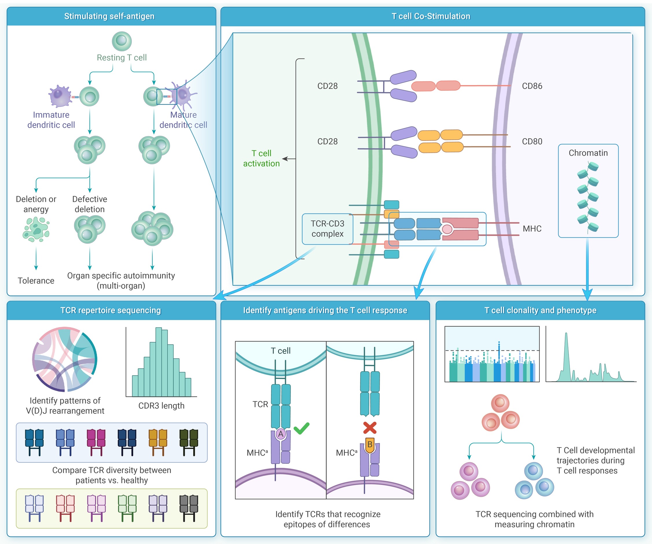

-
Figure 1.
Literature screening workflow.
-
Figure 2.
ML model architectures and data processing workflows.
-
Figure 3.
Schematic diagram of the mechanisms of TCR-mediated immune responses.
-
Figure 4.
Schematic diagram of ML-driven antibody development process.
