
Neural regulation in cancer: A comprehensive review from tumorigenesis and progression to immune evasion
-
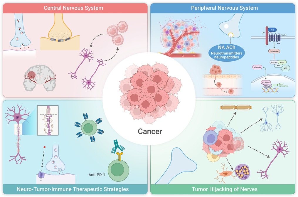
GRAPHICAL ABSTRACT
 DownLoad:
Full size image
DownLoad:
Full size image
-
PUBLIC SUMMARY
-
Nerves actively shape how cancers grow, spread, and evade immune attack.
Tumors rewire neural circuits and exploit neurotransmitters to create a supportive niche.
Neural signals alter immune cells, angiogenesis, metabolism, and treatment response.
Targeting neuro–tumor–immune pathways may enable new combination cancer therapies.
-
-
ABSTRACT
Scientific tradition long treated neural and immune activities as essentially separate domains. Contrary to this historical view, studies from the past several decades have fundamentally transformed our understanding. Across various cancers, the nervous system establishes a dynamic, bidirectional regulatory network connecting neural elements, tumors, and immune cells. This occurs through direct interactions (including paracrine communication and electrochemical neuron-cancer signaling) and indirect mechanisms involving the tumor microenvironment (TME) regulation. A two-way regulatory relationship characterizes the nervous system-tumor interaction, combining neural control of oncogenesis with tumor-driven neural plasticity to form a pathological feedback loop. Our analysis delves into how the central nervous system (CNS) and the peripheral nervous system (PNS) directly regulate tumor behavior and anti-tumor immunity, and how cancers hijack normal neural signaling pathways to promote growth and immune evasion. We further examine how tumors and anticancer treatments reciprocally affect neural functionality and immune homeostasis. -

-
REFERENCES
[1] Baraldi J.H., Martyn G.V., Shurin G.V., et al. (2022). Tumor innervation: History, methodologies, and significance. Cancers 14:1979. DOI:10.3390/cancers14081979 [2] Magnon C., Hall S.J., Lin J., et al. (2013). Autonomic nerve development contributes to prostate cancer progression. Science 341:1236361. DOI:10.1126/science.1236361 [3] Wang W., Li L., Chen N., et al. (2020). Nerves in the tumor microenvironment: Origin and effects. Front. Cell Dev. Biol. 8:601738. DOI:10.3389/fcell.2020.601738 [4] Yaman I., Ağaç Çobanoğlu D., Xie T., et al. (2022). Advances in understanding cancer-associated neurogenesis and its implications on the neuroimmune axis in cancer. Pharmacol. Ther. 239:108199. DOI:10.1016/j.pharmthera.2022.108199 [5] O’Donnell M., Chance R.K. and Bashaw G.J. (2009). Axon growth and guidance: Receptor regulation and signal transduction. Annu. Rev. Neurosci. 32:383−412. DOI:10.1146/annurev.neuro.051508.135614 [6] Kamiya A., Hiyama T., Fujimura A., et al. (2021). Sympathetic and parasympathetic innervation in cancer: Therapeutic implications. Clin. Auton. Res. 31:165−178. DOI:10.1007/s10286-020-00724-y [7] Mancusi R. and Monje M. (2023). The neuroscience of cancer. Nature 618:467−479. DOI:10.1038/s41586-023-05968-y [8] Sharkey K.A. and Mawe G.M. (2023). The enteric nervous system. Physiol. Rev. 103:1487−1564. DOI:10.1152/physrev.00018.2022 [9] Le T.T. and Oudin M.J. (2023). Understanding and modeling nerve-cancer interactions. Dis. Model. Mech. 16:dmm049729. DOI:10.1242/dmm.049729 [10] Colombo E. and Farina C. (2016). Astrocytes: Key regulators of neuroinflammation. Trends Immunol. 37:608−620. DOI:10.1016/j.it.2016.06.006 [11] Christopherson K.S., Ullian E.M., Stokes C.C.A., et al. (2005). Thrombospondins are astrocyte-secreted proteins that promote CNS synaptogenesis. Cell 120:421−433. DOI:10.1016/j.cell.2004.12.020 [12] Mauch D.H., Nägler K., Schumacher S., et al. (2001). CNS synaptogenesis promoted by glia-derived cholesterol. Science 294:1354−1357. DOI:10.1126/science.294.5545.1354 [13] Hughes E.G., Kang S.H., Fukaya M., et al. (2013). Oligodendrocyte progenitors balance growth with self-repulsion to achieve homeostasis in the adult brain. Nat. Neurosci. 16:668−676. DOI:10.1038/nn.3390 [14] Pan Y. and Monje M. (2020). Activity shapes neural circuit form and function: A historical perspective. J. Neurosci. 40:944−954. DOI:10.1523/JNEUROSCI.0740-19.2019 [15] Winkler F., Venkatesh H.S., Amit M., et al. (2023). Cancer neuroscience: State of the field, emerging directions. Cell 186:1689−1707. DOI:10.1016/j.cell.2023.02.002 [16] Varadarajan S.G., Hunyara J.L., Hamilton N.R., et al. (2022). Central nervous system regeneration. Cell 185:77−94. DOI:10.1016/j.cell.2021.10.029 [17] Venkatesh H.S., Morishita W., Geraghty A.C., et al. (2019). Electrical and synaptic integration of glioma into neural circuits. Nature 573:539−545. DOI:10.1038/s41586-019-1563-y [18] Venkataramani V., Tanev D.I., Strahle C., et al. (2019). Glutamatergic synaptic input to glioma cells drives brain tumour progression. Nature 573:532−538. DOI:10.1038/s41586-019-1564-x [19] Lan Y., Zou S., Wang W., et al. (2023). Progress in cancer neuroscience. MedComm 4:e431. DOI:10.1002/mco2.431 [20] Taylor K.R., Barron T., Hui A., et al. (2023). Glioma synapses recruit mechanisms of adaptive plasticity. Nature 623:366−374. DOI:10.1038/s41586-023-06678-1 [21] Pan Y., Hysinger J.D., Barron T., et al. (2021). NF1 mutation drives neuronal activity-dependent initiation of optic glioma. Nature 594:277−282. DOI:10.1038/s41586-021-03580-6 [22] Chen P., Wang W., Liu R., et al. (2022). Olfactory sensory experience regulates gliomagenesis via neuronal IGF1. Nature 606:550−556. DOI:10.1038/s41586-022-04719-9 [23] Zeng Q., Michael I.P., Zhang P., et al. (2019). Synaptic proximity enables NMDAR signalling to promote brain metastasis. Nature 573:526−531. DOI:10.1038/s41586-019-1576-6 [24] Sakthivelu V., Schmitt A., Odenthal F., et al. (2025). Functional synapses between neurons and small cell lung cancer. Nature 646:1243−1253. DOI:10.1038/s41586-025-09434-9 [25] Wu C. and Sun D. (2015). GABA receptors in brain development, function, and injury. Metab. Brain Dis. 30:367−379. DOI:10.1007/s11011-014-9560-1 [26] Hills K.E., Kostarelos K. and Wykes R.C. (2022). Converging mechanisms of epileptogenesis and their insight in glioblastoma. Front. Mol. Neurosci. 15:903115. DOI:10.3389/fnmol.2022.903115 [27] Labrakakis C., Patt S., Hartmann J., et al. (1998). Functional GABA(a) receptors on human glioma cells. Eur. J. Neurosci. 10:231−238. DOI:10.1046/j.1460-9568.1998.00036.x [28] Campbell S.L., Robel S., Cuddapah V.A., et al. (2015). GABAergic disinhibition and impaired KCC2 cotransporter activity underlie tumor-associated epilepsy. Glia 63:23−36. DOI:10.1002/glia.22730 [29] Pallud J., Le Van Quyen M., Bielle F., et al. (2014). Cortical GABAergic excitation contributes to epileptic activities around human glioma. Sci. Transl. Med. 6:244ra89. DOI:10.1126/scitranslmed.3008065 [30] Müller-Längle A., Lutz H., Hehlgans S., et al. (2019). NMDA receptor-mediated signaling pathways enhance radiation resistance, survival and migration in glioblastoma cells-a potential target for adjuvant radiotherapy. Cancers 11:503. DOI:10.3390/cancers11040503 [31] Osswald M., Solecki G., Wick W., et al. (2016). A malignant cellular network in gliomas: Potential clinical implications. Neuro Oncol. 18:479−485. DOI:10.1093/neuonc/now014 [32] Osswald M., Jung E., Sahm F., et al. (2015). Brain tumour cells interconnect to a functional and resistant network. Nature 528:93−98. DOI:10.1038/nature16071 [33] Venkataramani V., Yang Y., Schubert M.C., et al. (2022). Glioblastoma hijacks neuronal mechanisms for brain invasion. Cell 185:2899−2917.e31. DOI:10.1016/j.cell.2022.06.054 [34] Guo Y., Li Y., Su P., et al. (2025). Tumor microtubes: A new potential therapeutic target for high-grade gliomas. J. Neuropathol. Exp. Neurol. 84:93−103. DOI:10.1093/jnen/nlae119 [35] Watson D.C., Bayik D., Storevik S., et al. (2023). GAP43-dependent mitochondria transfer from astrocytes enhances glioblastoma tumorigenicity. Nat. Cancer 4:648−664. DOI:10.1038/s43018-023-00556-5 [36] Alshehri R.S., Alrawaili M.S., Zawawi B.M.H., et al. (2025). Pathophysiology of status epilepticus revisited. Int. J. Mol. Sci. 26:7502. DOI:10.3390/ijms26157502 [37] Sontheimer H. (2008). A role for glutamate in growth and invasion of primary brain tumors. J. Neurochem. 105:287−295. DOI:10.1111/j.1471-4159.2008.05301.x [38] Ishibashi K., Ichinose T., Kadokawa R., et al. (2024). Astrocyte-induced mGluR1 activates human lung cancer brain metastasis via glutamate-dependent stabilization of EGFR. Dev. Cell 59:579−594.e6. DOI:10.1016/j.devcel.2024.01.010 [39] Hujber Z., Horváth G., Petővári G., et al. (2018). GABA, glutamine, glutamate oxidation and succinic semialdehyde dehydrogenase expression in human gliomas. J. Exp. Clin. Cancer Res. 37:271. DOI:10.1186/s13046-018-0946-5 [40] Li N., Xu X., Liu D., et al. (2023). The delta subunit of the GABAA receptor is necessary for the GPT2-promoted breast cancer metastasis. Theranostics 13:1355−1369. DOI:10.7150/thno.80544 [41] Azuma H., Inamoto T., Sakamoto T., et al. (2003). Gamma-aminobutyric acid as a promoting factor of cancer metastasis; induction of matrix metalloproteinase production is potentially its underlying mechanism. Cancer Res. 63:8090−8096. [42] Moreno-Smith M., Lu C., Shahzad M.M.K., et al. (2011). Dopamine blocks stress-mediated ovarian carcinoma growth. Clin. Cancer Res. 17:3649−3659. DOI:10.1158/1078-0432.CCR-10-2441 [43] Caragher S.P., Hall R.R., Ahsan R., et al. (2018). Monoamines in glioblastoma: Complex biology with therapeutic potential. Neuro Oncol. 20:1014−1025. DOI:10.1093/neuonc/nox210 [44] He Y., Li J., Koga T., et al. (2020). Epidermal growth factor receptor as a molecular determinant of glioblastoma response to dopamine receptor D2 inhibitors. Neuro Oncol. 23:400−411. DOI:10.1093/neuonc/noaa188 [45] Wang Y., Wang X., Wang K., et al. (2023). Chronic stress accelerates glioblastoma progression via DRD2/ERK/β-catenin axis and dopamine/ERK/TH positive feedback loop. J. Exp. Clin. Cancer Res. 42:161. DOI:10.1186/s13046-023-02728-8 [46] Luikart B.W., Nef S., Virmani T., et al. (2005). TrkB has a cell-autonomous role in the establishment of hippocampal Schaffer collateral synapses. J. Neurosci. 25:3774−3786. DOI:10.1523/JNEUROSCI.0041-05.2005 [47] Chapleau C.A. and Pozzo-Miller L. (2012). Divergent roles of p75NTR and Trk receptors in BDNF’s effects on dendritic spine density and morphology. Neural Plast. 2012:578057. DOI:10.1155/2012/578057 [48] Nakata H. and Nakamura S. (2007). Brain-derived neurotrophic factor regulates AMPA receptor trafficking to post-synaptic densities via IP3R and TRPC calcium signaling. FEBS Lett. 581:2047−2054. DOI:10.1016/j.febslet.2007.04.041 [49] Li X. and Wolf M.E. (2011). Brain-derived neurotrophic factor rapidly increases AMPA receptor surface expression in rat nucleus accumbens. Eur. J. Neurosci. 34:190−198. DOI:10.1111/j.1460-9568.2011.07754.x [50] Figurov A., Pozzo-Miller L.D., Olafsson P., et al. (1996). Regulation of synaptic responses to high-frequency stimulation and LTP by neurotrophins in the hippocampus. Nature 381:706−709. DOI:10.1038/381706a0 [51] Pan Y. and Monje M. (2022). Neuron-glial interactions in health and brain cancer. Adv. Biol. 6:e2200122. DOI:10.1002/adbi.202200122 [52] Venkatesh H.S., Johung T.B., Caretti V., et al. (2015). Neuronal activity promotes glioma growth through neuroligin-3 secretion. Cell 161:803−816. DOI:10.1016/j.cell.2015.04.012 [53] Bulstrode H., Jones L.M., Siney E.J., et al. (2012). A-disintegrin and metalloprotease (ADAM) 10 and 17 promote self-renewal of brain tumor sphere forming cells. Cancer Lett. 326:79−87. DOI:10.1016/j.canlet.2012.07.022 [54] Kohutek Z.A., diPierro C.G., Redpath G.T., et al. (2009). ADAM-10-mediated N-cadherin cleavage is protein kinase C-α dependent and promotes glioblastoma cell migration. J. Neurosci. 29:4605−4615. DOI:10.1523/JNEUROSCI.5126-08.2009 [55] Venkatesh H.S., Tam L.T., Woo P.J., et al. (2017). Targeting neuronal activity-regulated neuroligin-3 dependency in high-grade glioma. Nature 549:533−537. DOI:10.1038/nature24014 [56] Liu R., Qin X.P., Zhuang Y., et al. (2018). Glioblastoma recurrence correlates with NLGN3 levels. Cancer Med. 7:2848−2859. DOI:10.1002/cam4.1538 [57] Yun E.J., Kim D., Kim S., et al. (2023). Targeting Wnt/β-catenin-mediated upregulation of oncogenic NLGN3 suppresses cancer stem cells in glioblastoma. Cell Death Dis. 14:423. DOI:10.1038/s41419-023-05967-x [58] Xue C., Chu Q., Shi Q., et al. (2025). Wnt signaling pathways in biology and disease: Mechanisms and therapeutic advances. Signal Transduct. Target. Ther. 10:106. DOI:10.1038/s41392-025-02142-w [59] Filippou P.S., Karagiannis G.S. and Constantinidou A. (2020). Midkine (MDK) growth factor: A key player in cancer progression and a promising therapeutic target. Oncogene 39:2040−2054. DOI:10.1038/s41388-019-1124-8 [60] Daginakatte G.C. and Gutmann D.H. (2007). Neurofibromatosis-1 (Nf1) heterozygous brain microglia elaborate paracrine factors that promote Nf1-deficient astrocyte and glioma growth. Hum. Mol. Genet. 16:1098−1112. DOI:10.1093/hmg/ddm059 [61] Solga A.C., Pong W.W., Kim K.Y., et al. (2015). RNA sequencing of tumor-associated microglia reveals Ccl5 as a stromal chemokine critical for neurofibromatosis-1 glioma growth. Neoplasia 17:776−788. DOI:10.1016/j.neo.2015.10.002 [62] Pan Y., Smithson L.J., Ma Y., et al. (2017). Ccl5 establishes an autocrine high-grade glioma growth regulatory circuit critical for mesenchymal glioblastoma survival. Oncotarget 8:32977−32989. DOI:10.18632/oncotarget.16516 [63] Guo X., Pan Y. and Gutmann D.H. (2019). Genetic and genomic alterations differentially dictate low-grade glioma growth through cancer stem cell-specific chemokine recruitment of T cells and microglia. Neuro Oncol. 21:1250−1262. DOI:10.1093/neuonc/noz080 [64] Bleakley L.E. and Reid C.A. (2024). HCN1 epilepsy: From genetics and mechanisms to precision therapies. J. Neurochem. 168:3891−3910. DOI:10.1111/jnc.15928 [65] Song K.W., Lim M. and Monje M. (2025). Complex neural-immune interactions shape glioma immunotherapy. Immunity 58:1140−1160. DOI:10.1016/j.immuni.2025.04.017 [66] Iadecola C. and Anrather J. (2011). The immunology of stroke: From mechanisms to translation. Nat. Med. 17:796−808. DOI:10.1038/nm.2399 [67] Brown C.E., Starr R., Aguilar B., et al. (2012). Stem-like tumor-initiating cells isolated from IL13Rα2 expressing gliomas are targeted and killed by IL13-zetakine-redirected T cells. Clin. Cancer Res. 18:2199−2209. DOI:10.1158/1078-0432.CCR-11-1669 [68] Flores C., Wildes T., Dean B.D., et al. (2019). Massive clonal expansion of medulloblastoma-specific T cells during adoptive cellular therapy. Sci. Adv. 5:eaav9879. DOI:10.1126/sciadv.aav9879 [69] Murad S., Michen S., Becker A., et al. (2022). NKG2C+ NK cells for immunotherapy of glioblastoma multiforme. Int. J. Mol. Sci. 23:5857. DOI:10.3390/ijms23105857 [70] Reardon D.A., Gokhale P.C., Klein S.R., et al. (2016). Glioblastoma eradication following immune checkpoint blockade in an orthotopic, immunocompetent model. Cancer Immunol. Res. 4:124−135. DOI:10.1158/2326-6066.CIR-15-0151 [71] Hoang-Minh L.B. and Mitchell D.A. (2018). Immunotherapy for brain tumors. Curr. Treat. Options Oncol. 19:60. DOI:10.1007/s11864-018-0576-3 [72] Khanmammadova N., Islam S., Sharma P., et al. (2023). Neuro-immune interactions and immuno-oncology. Trends Cancer 9:636−649. DOI:10.1016/j.trecan.2023.05.002 [73] Sampson J.H., Gunn M.D., Fecci P.E., et al. (2020). Brain immunology and immunotherapy in brain tumours. Nat. Rev. Cancer 20:12−25. DOI:10.1038/s41568-019-0224-7 [74] Linares C.A., Varghese A., Ghose A., et al. (2023). Hallmarks of the tumour microenvironment of gliomas and its interaction with emerging immunotherapy modalities. Int. J. Mol. Sci. 24:13215. DOI:10.3390/ijms241713215 [75] Rustenhoven J. (2021). A privileged brain. Science 374:548. DOI:10.1126/science.abl7122 [76] Mazzitelli J.A., Smyth L.C.D., Cross K.A., et al. (2022). Cerebrospinal fluid regulates skull bone marrow niches via direct access through dural channels. Nat. Neurosci. 25:555−560. DOI:10.1038/s41593-022-01029-1 [77] Pulous F.E., Cruz-Hernández J.C., Yang C., et al. (2022). Cerebrospinal fluid can exit into the skull bone marrow and instruct cranial hematopoiesis in mice with bacterial meningitis. Nat. Neurosci. 25:567−576. DOI:10.1038/s41593-022-01060-2 [78] Ringstad G. and Eide P.K. (2022). Molecular trans-dural efflux to skull bone marrow in humans with CSF disorders. Brain 145:1464−1472. DOI:10.1093/brain/awab388 [79] Wang Y., Chen D., Xu D., et al. (2021). Early developing B cells undergo negative selection by central nervous system-specific antigens in the meninges. Immunity 54:2784−2794.e6. DOI:10.1016/j.immuni.2021.09.016 [80] Rustenhoven J., Drieu A., Mamuladze T., et al. (2021). Functional characterization of the dural sinuses as a neuroimmune interface. Cell 184:1000−1016.e27. DOI:10.1016/j.cell.2020.12.040 [81] Shechter R., London A. and Schwartz M. (2013). Orchestrated leukocyte recruitment to immune-privileged sites: Absolute barriers versus educational gates. Nat. Rev. Immunol. 13:206−218. DOI:10.1038/nri3391 [82] Rustenhoven J. and Kipnis J. (2022). Brain borders at the central stage of neuroimmunology. Nature 612:417−429. DOI:10.1038/s41586-022-05474-7 [83] Hu X., Deng Q., Ma L., et al. (2020). Meningeal lymphatic vessels regulate brain tumor drainage and immunity. Cell Res. 30:229−243. DOI:10.1038/s41422-020-0287-8 [84] Weller R.O., Djuanda E., Yow H.Y., et al. (2009). Lymphatic drainage of the brain and the pathophysiology of neurological disease. Acta Neuropathol. 117:1−14. DOI:10.1007/s00401-008-0457-0 [85] Swartz M.A. (2014). Immunomodulatory roles of lymphatic vessels in cancer progression. Cancer Immunol. Res. 2:701−707. DOI:10.1158/2326-6066.CIR-14-0115 [86] Fransen M.F., Schoonderwoerd M., Knopf P., et al. (2018). Tumor-draining lymph nodes are pivotal in PD-1/PD-L1 checkpoint therapy. JCI Insight 3:e124507. DOI:10.1172/jci.insight.124507 [87] Song E., Mao T., Dong H., et al. (2020). VEGF-C-driven lymphatic drainage enables immunosurveillance of brain tumours. Nature 577:689−694. DOI:10.1038/s41586-019-1912-x [88] Schiller M., Ben-Shaanan T.L. and Rolls A. (2021). Neuronal regulation of immunity: Why, how and where. Nat. Rev. Immunol. 21:20−36. DOI:10.1038/s41577-020-0387-1 [89] Ganong W.F. (2000). Circumventricular organs: Definition and role in the regulation of endocrine and autonomic function. Clin. Exp. Pharmacol. Physiol. 27:422−427. DOI:10.1046/j.1440-1681.2000.03259.x [90] Manore S.G., Doheny D.L., Wong G.L., et al. (2022). IL-6/JAK/STAT3 signaling in breast cancer metastasis: Biology and treatment. Front. Oncol. 12:866014. DOI:10.3389/fonc.2022.866014 [91] Magnon C. and Hondermarck H. (2023). The neural addiction of cancer. Nat. Rev. Cancer 23:317−334. DOI:10.1038/s41568-023-00556-8 [92] Cerqueira J.J., Mailliet F., Almeida O.F.X., et al. (2007). The prefrontal cortex as a key target of the maladaptive response to stress. J. Neurosci. 27:2781−2787. DOI:10.1523/JNEUROSCI.4372-06.2007 [93] Liston C., Miller M.M., Goldwater D.S., et al. (2006). Stress-induced alterations in prefrontal cortical dendritic morphology predict selective impairments in perceptual attentional set-shifting. J. Neurosci. 26:7870−7874. DOI:10.1523/JNEUROSCI.1184-06.2006 [94] Hare B.D., Shinohara R., Liu R.J., et al. (2019). Optogenetic stimulation of medial prefrontal cortex Drd1 neurons produces rapid and long-lasting antidepressant effects. Nat. Commun. 10:223. DOI:10.1038/s41467-018-08168-9 [95] Surman M. and Janik M.E. (2017). Stress and its molecular consequences in cancer progression. Postepy Hig. Med. Dosw. 71:485−499. DOI:10.5604/01.3001.0010.3830 [96] Balood M., Ahmadi M., Eichwald T., et al. (2022). Nociceptor neurons affect cancer immunosurveillance. Nature 611:405−412. DOI:10.1038/s41586-022-05374-w [97] Xu X.R., Xiao Q., Hong Y.C., et al. (2021). Activation of dopaminergic VTA inputs to the mPFC ameliorates chronic stress-induced breast tumor progression. CNS Neurosci. Ther. 27:206−219. DOI:10.1111/cns.13465 [98] Li Y., Yu H., Li Z.M., et al. (2025). Colorectal cancer cells hijack a brain-gut polysynaptic circuit from the lateral septum to enteric neurons to sustain tumor growth. Nat. Cancer 6:1800−1820. DOI:10.1038/s43018-025-01033-x [99] Hanahan D. and Weinberg R.A. (2000). The hallmarks of cancer. Cell 100:57−70. DOI:10.1016/S0092-8674(00)81683-9 [100] Hanahan D. (2022). Hallmarks of cancer: New dimensions. Cancer Discov. 12:31−46. DOI:10.1158/2159-8290.CD-21-1059 [101] Knighton D., Ausprunk D., Tapper D., et al. (1977). Avascular and vascular phases of tumour growth in the chick embryo. Br. J. Cancer 35:347−356. DOI:10.1038/bjc.1977.49 [102] Saloman J.L., Albers K.M., Rhim A.D., et al. (2016). Can stopping nerves, stop cancer. Trends Neurosci. 39:880−889. DOI:10.1016/j.tins.2016.10.002 [103] Vázquez-Prado J., Bracho-Valdés I., Cervantes-Villagrana R.D., et al. (2016). Gβγ pathways in cell polarity and migration linked to oncogenic GPCR signaling: Potential relevance in tumor microenvironment. Mol. Pharmacol. 90:573−586. DOI:10.1124/mol.116.105338 [104] Cervantes-Villagrana R.D., Albores-García D., Cervantes-Villagrana A.R., et al. (2020). Tumor-induced neurogenesis and immune evasion as targets of innovative anti-cancer therapies. Signal Transduct. Target. Ther. 5:99. DOI:10.1038/s41392-020-0205-z [105] Lu X., Cheng L., Yang C., et al. (2024). Crosstalk between bladder cancer and the tumor microenvironment: Molecular mechanisms and targeted therapy. Innov. Med. 2:100094. DOI:10.59717/j.xinn-med.2024.100094 [106] Fan R., Lu X., Xu C., et al. (2025). Multidimensional regulation of the tumor microenvironment. Innov. Med. 3:100166. DOI:10.59717/j.xinn-med.2025.100166 [107] Xia Y., Wei Y., Li Z.Y., et al. (2019). Catecholamines contribute to the neovascularization of lung cancer via tumor-associated macrophages. Brain Behav. Immun. 81:111−121. DOI:10.1016/j.bbi.2019.06.004 [108] Beider K., Bitner H., Leiba M., et al. (2014). Multiple myeloma cells recruit tumor-supportive macrophages through the CXCR4/CXCL12 axis and promote their polarization toward the M2 phenotype. Oncotarget 5:11283−11296. DOI:10.18632/oncotarget.2207 [109] Sinha P., Clements V.K., Fulton A.M., et al. (2007). Prostaglandin E2 promotes tumor progression by inducing myeloid-derived suppressor cells. Cancer Res. 67:4507−4513. DOI:10.1158/0008-5472.CAN-06-4174 [110] Wu J., Gao F.X., Wang C., et al. (2019). IL-6 and IL-8 secreted by tumour cells impair the function of NK cells via the STAT3 pathway in oesophageal squamous cell carcinoma. J. Exp. Clin. Cancer Res. 38:321. DOI:10.1186/s13046-019-1310-0 [111] Shurin G.V., Vats K., Kruglov O., et al. (2022). Tumor-induced T cell polarization by Schwann cells. Cells 11:3541. DOI:10.3390/cells11223541 [112] Baruch E.N., Gleber-Netto F.O., Nagarajan P., et al. (2025). Cancer-induced nerve injury promotes resistance to anti-PD-1 therapy. Nature 646:462−473. DOI:10.1038/s41586-025-09370-8 [113] Rotshenker S. (2011). Wallerian degeneration: The innate-immune response to traumatic nerve injury. J. Neuroinflammation 8:109. DOI:10.1186/1742-2094-8-109 [114] Zhou X., Zhang B., Hu J., et al. (2024). Igniting cold tumors of intrahepatic cholangiocarcinoma: An insight into immune evasion and tumor immune microenvironment. Innov. Med. 2:100052. DOI:10.59717/j.xinn-med.2024.100052 [115] Zahalka A.H., Arnal-Estapé A., Maryanovich M., et al. (2017). Adrenergic nerves activate an angio-metabolic switch in prostate cancer. Science 358:321−326. DOI:10.1126/science.aah5072 [116] Sierra J.R., Corso S., Caione L., et al. (2008). Tumor angiogenesis and progression are enhanced by Sema4D produced by tumor-associated macrophages. J. Exp. Med. 205:1673−1685. DOI:10.1084/jem.20072602 [117] Garrido M.P., Torres I., Vega M., et al. (2019). Angiogenesis in gynecological cancers: Role of neurotrophins. Front. Oncol. 9:913. DOI:10.3389/fonc.2019.00913 [118] Caterina M.J., Schumacher M.A., Tominaga M., et al. (1997). The capsaicin receptor: A heat-activated ion channel in the pain pathway. Nature 389:816−824. DOI:10.1038/39807 [119] Caterina M.J., Leffler A., Malmberg A.B., et al. (2000). Impaired nociception and pain sensation in mice lacking the capsaicin receptor. Science 288:306−313. DOI:10.1126/science.288.5464.306 [120] Davis J.B., Gray J., Gunthorpe M.J., et al. (2000). Vanilloid receptor-1 is essential for inflammatory thermal hyperalgesia. Nature 405:183−187. DOI:10.1038/35012076 [121] Abraira V.E. and Ginty D.D. (2013). The sensory neurons of touch. Neuron 79:618−639. DOI:10.1016/j.neuron.2013.07.051 [122] Blake K.J., Baral P., Voisin T., et al. (2018). Staphylococcus aureus produces pain through pore-forming toxins and neuronal TRPV1 that is silenced by QX-314. Nat. Commun. 9:37. DOI:10.1038/s41467-017-02448-6 [123] Chiu I.M., von Hehn C.A. and Woolf C.J. (2012). Neurogenic inflammation and the peripheral nervous system in host defense and immunopathology. Nat. Neurosci. 15:1063−1067. DOI:10.1038/nn.3144 [124] Saloman J.L., Albers K.M., Li D., et al. (2016). Ablation of sensory neurons in a genetic model of pancreatic ductal adenocarcinoma slows initiation and progression of cancer. Proc. Natl. Acad. Sci. USA 113:3078−3083. DOI:10.1073/pnas.1512603113 [125] Restaino A.C., Walz A., Vermeer S.J., et al. (2023). Functional neuronal circuits promote disease progression in cancer. Sci. Adv. 9:eade4443. DOI:10.1126/sciadv.ade4443 [126] Navarro X., Verdú E., Wendelscafer-Crabb G., et al. (1995). Innervation of cutaneous structures in the mouse hind paw: A confocal microscopy immunohistochemical study. J. Neurosci. Res. 41:111−120. DOI:10.1002/jnr.490410113 [127] Ishida-Yamamoto A., Senba E. and Tohyama M. (1989). Distribution and fine structure of calcitonin gene-related peptide-like immunoreactive nerve fibers in the rat skin. Brain Res. 491:93−101. DOI:10.1016/0006-8993(89)90090-5 [128] Pundavela J., Demont Y., Jobling P., et al. (2014). ProNGF correlates with Gleason score and is a potential driver of nerve infiltration in prostate cancer. Am. J. Pathol. 184:3156−3162. DOI:10.1016/j.ajpath.2014.08.009 [129] Crosson T., Roversi K., Balood M., et al. (2019). Profiling of how nociceptor neurons detect danger - new and old foes. J. Intern. Med. 286:268−289. DOI:10.1111/joim.12957 [130] Crosson T., Wang J.C., Doyle B., et al. (2021). FcεR1-expressing nociceptors trigger allergic airway inflammation. J. Allergy Clin. Immunol. 147:2330−2342. DOI:10.1016/j.jaci.2020.12.644 [131] Shalabi S., Belayachi A. and Larrivée B. (2024). Involvement of neuronal factors in tumor angiogenesis and the shaping of the cancer microenvironment. Front. Immunol. 15:1284629. DOI:10.3389/fimmu.2024.1284629 [132] Olofsson P.S., Steinberg B.E., Sobbi R., et al. (2016). Blood pressure regulation by CD4(+) lymphocytes expressing choline acetyltransferase. Nat. Biotechnol. 34:1066−1071. DOI:10.1038/nbt.3663 [133] Budczies J., Pfitzner B.M., Györffy B., et al. (2015). Glutamate enrichment as new diagnostic opportunity in breast cancer. Int. J. Cancer 136:1619−1628. DOI:10.1002/ijc.29152 [134] Yu L.J., Wall B.A., Wangari-Talbot J., et al. (2017). Metabotropic glutamate receptors in cancer. Neuropharmacology 115:193−202. DOI:10.1016/j.neuropharm.2016.02.011 [135] Wang J.B., Erickson J.W., Fuji R., et al. (2010). Targeting mitochondrial glutaminase activity inhibits oncogenic transformation. Cancer Cell 18:207−219. DOI:10.1016/j.ccr.2010.08.009 [136] Huang F., Zhang Q., Ma H., et al. (2014). Expression of glutaminase is upregulated in colorectal cancer and of clinical significance. Int. J. Clin. Exp. Pathol. 7:1093−1100. [137] Seltzer M.J., Bennett B.D., Joshi A.D., et al. (2010). Inhibition of glutaminase preferentially slows growth of glioma cells with mutant IDH1. Cancer Res. 70:8981−8987. DOI:10.1158/0008-5472.Can-10-1666 [138] Son J., Lyssiotis C.A., Ying H., et al. (2013). Glutamine supports pancreatic cancer growth through a KRAS-regulated metabolic pathway. Nature 496:101−105. DOI:10.1038/nature12040 [139] Koochekpour S., Majumdar S., Azabdaftari G., et al. (2012). Serum glutamate levels correlate with Gleason score and glutamate blockade decreases proliferation, migration, and invasion and induces apoptosis in prostate cancer cells. Clin. Cancer Res. 18:5888−5901. DOI:10.1158/1078-0432.Ccr-12-1308 [140] Gross M.I., Demo S.D., Dennison J.B., et al. (2014). Antitumor activity of the glutaminase inhibitor CB-839 in triple-negative breast cancer. Mol. Cancer Ther. 13:890−901. DOI:10.1158/1535-7163.Mct-13-0870 [141] Yu T., Van der Jeught K., Zhu H., et al. (2025). Inhibition of glutamate-to-glutathione flux promotes tumor antigen presentation in colorectal cancer cells. Adv. Sci. 12:e2310308. DOI:10.1002/advs.202310308 [142] García-Gaytán A.C., Hernández-Abrego A., Díaz-Muñoz M., et al. (2022). Glutamatergic system components as potential biomarkers and therapeutic targets in cancer in non-neural organs. Front. Endocrinol. 13:1029210. DOI:10.3389/fendo.2022.1029210 [143] Stepulak A., Rola R., Polberg K., et al. (2014). Glutamate and its receptors in cancer. J. Neural Transm. 121:933−944. DOI:10.1007/s00702-014-1182-6 [144] Du J., Li X.H. and Li Y.J. (2016). Glutamate in peripheral organs: Biology and pharmacology. Eur. J. Pharmacol. 784:42−48. DOI:10.1016/j.ejphar.2016.05.009 [145] Martino J.J., Wall B.A., Mastrantoni E., et al. (2013). Metabotropic glutamate receptor 1 (Grm1) is an oncogene in epithelial cells. Oncogene 32:4366−4376. DOI:10.1038/onc.2012.471 [146] Liu J.W., Kim M.S., Nagpal J., et al. (2007). Quantitative hypermethylation of NMDAR2B in human gastric cancer. Int. J. Cancer 121:1994−2000. DOI:10.1002/ijc.22934 [147] Tamura H., Suzuki M., Moriya Y., et al. (2011). Aberrant methylation of N-methyl-D-aspartate receptor type 2B (NMDAR2B) in non-small cell carcinoma. BMC Cancer 11:220. DOI:10.1186/1471-2407-11-220 [148] Ren L., Liu C., Çifcibaşı K., et al. (2025). Sensory neurons drive pancreatic cancer progression through glutamatergic neuron-cancer pseudo-synapses. Cancer Cell 43:2241−2258.e8. DOI:10.1016/j.ccell.2025.09.003 [149] Herner A., Sauliunaite D., Michalski C.W., et al. (2011). Glutamate increases pancreatic cancer cell invasion and migration via AMPA receptor activation and Kras-MAPK signaling. Int. J. Cancer 129:2349−2359. DOI:10.1002/ijc.25898 [150] Mashaghi A., Marmalidou A., Tehrani M., et al. (2016). Neuropeptide substance P and the immune response. Cell Mol. Life Sci. 73:4249−4264. DOI:10.1007/s00018-016-2293-z [151] Manske J.M. and Hanson S.E. (2005). Substance-P-mediated immunomodulation of tumor growth in a murine model. Neuroimmunomodulation 12:201−210. DOI:10.1159/000085652 [152] Chernova I., Lai J.P., Li H., et al. (2009). Substance P (SP) enhances CCL5-induced chemotaxis and intracellular signaling in human monocytes, which express the truncated neurokinin-1 receptor (NK1R). J. Leukoc. Biol. 85:154−164. DOI:10.1189/jlb.0408260 [153] Coveñas R. and Muñoz M. (2022). Involvement of the substance P/neurokinin-1 receptor system in cancer. Cancers 14:3539. DOI:10.3390/cancers14143539 [154] Muñoz M., Rosso M., González-Ortega A., et al. (2010). The NK-1 receptor antagonist L-732,138 induces apoptosis and counteracts substance P-related mitogenesis in human melanoma cell lines. Cancers 2:611−623. DOI:10.3390/cancers2020611 [155] Garcia-Recio S., Fuster G., Fernandez-Nogueira P., et al. (2013). Substance P autocrine signaling contributes to persistent HER2 activation that drives malignant progression and drug resistance in breast cancer. Cancer Res. 73:6424−6434. DOI:10.1158/0008-5472.Can-12-4573 [156] Palma C., Nardelli F., Manzini S., et al. (1999). Substance P activates responses correlated with tumour growth in human glioma cell lines bearing tachykinin NK1 receptors. Br. J. Cancer 79:236−243. DOI:10.1038/sj.bjc.6690039 [157] Hong X., Ma J., Zheng S., et al. (2024). Advances in the research and application of neurokinin-1 receptor antagonists. J. Zhejiang Univ. Sci. B 25:91−105. DOI:10.1631/jzus.B2300455 [158] Coveñas R., Rodríguez F.D., Robinson P., et al. (2023). The repurposing of non-peptide neurokinin-1 receptor antagonists as antitumor drugs: An urgent challenge for aprepitant. Int. J. Mol. Sci. 24:15936. DOI:10.3390/ijms242115936 [159] Muñoz M., Crespo J.C., Crespo J.P., et al. (2019). Neurokinin-1 receptor antagonist aprepitant and radiotherapy, a successful combination therapy in a patient with lung cancer: A case report. Mol. Clin. Oncol. 11:50−54. DOI:10.3892/mco.2019.1857 [160] Nizam E., Köksoy S. and Erin N. (2020). NK1R antagonist decreases inflammation and metastasis of breast carcinoma cells metastasized to liver but not to brain; phenotype-dependent therapeutic and toxic consequences. Cancer Immunol. Immunother. 69:1639−1650. DOI:10.1007/s00262-020-02574-z [161] Holzmann B. (2013). Antiinflammatory activities of CGRP modulating innate immune responses in health and disease. Curr. Protein Pept. Sci. 14:268−274. DOI:10.2174/13892037113149990046 [162] Springer J., Geppetti P., Fischer A., et al. (2003). Calcitonin gene-related peptide as inflammatory mediator. Pulm. Pharmacol. Ther. 16:121−130. DOI:10.1016/s1094-5539(03)00049-x [163] Vats K., Kruglov O., Sahoo B., et al. (2022). Sensory nerves impede the formation of tertiary lymphoid structures and development of protective antimelanoma immune responses. Cancer Immunol. Res. 10:1141−1154. DOI:10.1158/2326-6066.Cir-22-0110 [164] McIlvried L.A., Atherton M.A., Horan N.L., et al. (2022). Sensory neurotransmitter calcitonin gene-related peptide modulates tumor growth and lymphocyte infiltration in oral squamous cell carcinoma. Adv. Biol. 6:e2200019. DOI:10.1002/adbi.202200019 [165] Toda M., Suzuki T., Hosono K., et al. (2008). Neuronal system-dependent facilitation of tumor angiogenesis and tumor growth by calcitonin gene-related peptide. Proc. Natl. Acad. Sci. USA 105:13550−13555. DOI:10.1073/pnas.0800767105 [166] Wang K., Ni B., Xie Y., et al. (2025). Nociceptor neurons promote PDAC progression and cancer pain by interaction with cancer-associated fibroblasts and suppression of natural killer cells. Cell Res. 35:362−380. DOI:10.1038/s41422-025-01098-4 [167] Zhi X., Wu F., Qian J., et al. (2025). Nociceptive neurons promote gastric tumour progression via a CGRP-RAMP1 axis. Nature 640:802−810. DOI:10.1038/s41586-025-08591-1 [168] Zhang Y., Lin C., Liu Z., et al. (2022). Cancer cells co-opt nociceptive nerves to thrive in nutrient-poor environments and upon nutrient-starvation therapies. Cell Metab. 34:1999−2017.e10. DOI:10.1016/j.cmet.2022.10.012 [169] Hou Y., Lin B., Xu T., et al. (2024). The neurotransmitter calcitonin gene-related peptide shapes an immunosuppressive microenvironment in medullary thyroid cancer. Nat. Commun. 15:5555. DOI:10.1038/s41467-024-49824-7 [170] Zhang H., Zhang Y., Zhou P., et al. (2025). Intra-tumoural RAMP1+ B cells promote resistance to neoadjuvant anti-PD-1-based therapy in oesophageal squamous cell carcinoma. Immunother. Adv. 5:ltaf012. DOI:10.1093/immadv/ltaf012 [171] Xiao Z., Wang J., Yang J., et al. (2025). Dendritic cells instruct T cell anti-tumor immunity and immunotherapy response. Innov. Med. 3:100128. DOI:10.59717/j.xinn-med.2025.100128 [172] Bujak J.K., Kosmala D., Szopa I.M., et al. (2019). Inflammation, cancer and immunity-implication of TRPV1 channel. Front. Oncol. 9:1087. DOI:10.3389/fonc.2019.01087 [173] Kiss F., Pohóczky K., Szállási A., et al. (2020). Transient receptor potential (TRP) channels in head-and-neck squamous cell carcinomas: Diagnostic, prognostic, and therapeutic potentials. Int. J. Mol. Sci. 21:6374. DOI:10.3390/ijms21176374 [174] Ravindranathan S., Passang T., Li J.M., et al. (2022). Targeting vasoactive intestinal peptide-mediated signaling enhances response to immune checkpoint therapy in pancreatic ductal adenocarcinoma. Nat. Commun. 13:6418. DOI:10.1038/s41467-022-34242-4 [175] Nance D.M. and Sanders V.M. (2007). Autonomic innervation and regulation of the immune system (1987-2007). Brain Behav. Immun. 21:736−745. DOI:10.1016/j.bbi.2007.03.008 [176] Rosas-Ballina M., Olofsson P.S., Ochani M., et al. (2011). Acetylcholine-synthesizing T cells relay neural signals in a vagus nerve circuit. Science 334:98−101. DOI:10.1126/science.1209985 [177] Sastry K.S., Karpova Y., Prokopovich S., et al. (2007). Epinephrine protects cancer cells from apoptosis via activation of cAMP-dependent protein kinase and BAD phosphorylation. J. Biol. Chem. 282:14094−14100. DOI:10.1074/jbc.M611370200 [178] Cole S.W., Nagaraja A.S., Lutgendorf S.K., et al. (2015). Sympathetic nervous system regulation of the tumour microenvironment. Nat. Rev. Cancer 15:563−572. DOI:10.1038/nrc3978 [179] Shan T., Ma Q., Zhang D., et al. (2011). β2-Adrenoceptor blocker synergizes with gemcitabine to inhibit the proliferation of pancreatic cancer cells via apoptosis induction. Eur. J. Pharmacol. 665:1−7. DOI:10.1016/j.ejphar.2011.04.055 [180] Zhang D., Ma Q.Y., Hu H.T., et al. (2010). β2-Adrenergic antagonists suppress pancreatic cancer cell invasion by inhibiting CREB, NFκB and AP-1. Cancer Biol. Ther. 10:19−29. DOI:10.4161/cbt.10.1.11944 [181] Allen J.K., Armaiz-Pena G.N., Nagaraja A.S., et al. (2018). Sustained adrenergic signaling promotes intratumoral innervation through BDNF induction. Cancer Res. 78:3233−3242. DOI:10.1158/0008-5472.Can-16-1701 [182] Globig A.M., Zhao S., Roginsky J., et al. (2023). The β(1)-adrenergic receptor links sympathetic nerves to T cell exhaustion. Nature 622:383−392. DOI:10.1038/s41586-023-06568-6 [183] Isaacs J.T. (2013). Cancer. Prostate cancer takes nerve. Science 341:134−135. DOI:10.1126/science.1241776 [184] Zhu J., Naulaerts S., Boudhan L., et al. (2023). Tumour immune rejection triggered by activation of α2-adrenergic receptors. Nature 618:607−615. DOI:10.1038/s41586-023-06110-8 [185] Sloan E.K., Priceman S.J., Cox B.F., et al. (2010). The sympathetic nervous system induces a metastatic switch in primary breast cancer. Cancer Res. 70:7042−7052. DOI:10.1158/0008-5472.Can-10-0522 [186] Podojil J.R. and Sanders V.M. (2003). Selective regulation of mature IgG1 transcription by CD86 and beta 2-adrenergic receptor stimulation. J. Immunol. 170:5143−5151. DOI:10.4049/jimmunol.170.10.5143 [187] Kohm A.P. and Sanders V.M. (2001). Norepinephrine and beta 2-adrenergic receptor stimulation regulate CD4+ T and B lymphocyte function in vitro and in vivo. Pharmacol. Rev. 53:487−525. [188] Kokolus K.M., Capitano M.L., Lee C.T., et al. (2013). Baseline tumor growth and immune control in laboratory mice are significantly influenced by subthermoneutral housing temperature. Proc. Natl. Acad. Sci. USA 110:20176−20181. DOI:10.1073/pnas.1304291110 [189] Kokolus K.M., Spangler H.M., Povinelli B.J., et al. (2014). Stressful presentations: Mild cold stress in laboratory mice influences phenotype of dendritic cells in naïve and tumor-bearing mice. Front. Immunol. 5:23. DOI:10.3389/fimmu.2014.00023 [190] Goldfarb Y., Sorski L., Benish M., et al. (2011). Improving postoperative immune status and resistance to cancer metastasis: A combined perioperative approach of immunostimulation and prevention of excessive surgical stress responses. Ann. Surg. 253:798−810. DOI:10.1097/SLA.0b013e318211d7b5 [191] Inbar S., Neeman E., Avraham R., et al. (2011). Do stress responses promote leukemia progression? An animal study suggesting a role for epinephrine and prostaglandin-E2 through reduced NK activity. PLoS One 6:e19246. DOI:10.1371/journal.pone.0019246. [192] Benish M., Bartal I., Goldfarb Y., et al. (2008). Perioperative use of beta-blockers and COX-2 inhibitors may improve immune competence and reduce the risk of tumor metastasis. Ann. Surg. Oncol. 15:2042−2052. DOI:10.1245/s10434-008-9890-5 [193] Mundy-Bosse B.L., Thornton L.M., Yang H.C., et al. (2011). Psychological stress is associated with altered levels of myeloid-derived suppressor cells in breast cancer patients. Cell Immunol. 270:80−87. DOI:10.1016/j.cellimm.2011.04.003 [194] Rosenne E., Sorski L., Shaashua L., et al. (2014). In vivo suppression of NK cell cytotoxicity by stress and surgery: Glucocorticoids have a minor role compared to catecholamines and prostaglandins. Brain Behav. Immun. 37:207−219. DOI:10.1016/j.bbi.2013.12.007 [195] Amit M., Takahashi H., Dragomir M.P., et al. (2020). Loss of p53 drives neuron reprogramming in head and neck cancer. Nature 578:449−454. DOI:10.1038/s41586-020-1996-3 [196] Mauffrey P., Tchitchek N., Barroca V., et al. (2019). Progenitors from the central nervous system drive neurogenesis in cancer. Nature 569:672−678. DOI:10.1038/s41586-019-1219-y [197] Kennedy O.J., Kicinski M., Valpione S., et al. (2022). Prognostic and predictive value of β-blockers in the EORTC 1325/KEYNOTE-054 phase III trial of pembrolizumab versus placebo in resected high-risk stage III melanoma. Eur. J. Cancer 165:97−112. DOI:10.1016/j.ejca.2022.01.017 [198] Qiao G., Chen M., Mohammadpour H., et al. (2021). Chronic adrenergic stress contributes to metabolic dysfunction and an exhausted phenotype in T cells in the tumor microenvironment. Cancer Immunol. Res. 9:651−664. DOI:10.1158/2326-6066.Cir-20-0445 [199] Zhou H., Shi B., Jia Y., et al. (2018). Expression and significance of autonomic nerves and α9 nicotinic acetylcholine receptor in colorectal cancer. Mol. Med. Rep. 17:8423−8431. DOI:10.3892/mmr.2018.8883 [200] Hicks B.M., Murray L.J., Powe D.G., et al. (2013). β-Blocker usage and colorectal cancer mortality: A nested case-control study in the UK Clinical Practice Research Datalink cohort. Ann. Oncol. 24:3100−3106. DOI:10.1093/annonc/mdt381 [201] Nissen M.D., Sloan E.K. and Mattarollo S.R. (2018). β-Adrenergic signaling impairs antitumor CD8(+) T-cell responses to B-cell lymphoma immunotherapy. Cancer Immunol. Res. 6:98−109. DOI:10.1158/2326-6066.Cir-17-0401 [202] Brem S. (2024). Vagus nerve stimulation: Novel concept for the treatment of glioblastoma and solid cancers by cytokine (interleukin-6) reduction, attenuating the SASP, enhancing tumor immunity. Brain Behav. Immun. Health 42:100859. DOI:10.1016/j.bbih.2024.100859 [203] Cortese N., Rigamonti A., Mantovani A., et al. (2020). The neuro-immune axis in cancer: Relevance of the peripheral nervous system to the disease. Immunol. Lett. 227:60−65. DOI:10.1016/j.imlet.2020.07.010 [204] Hutchings C., Phillips J.A. and Djamgoz M.B.A. (2020). Nerve input to tumours: Pathophysiological consequences of a dynamic relationship. Biochim. Biophys. Acta Rev. Cancer 1874:188411. DOI:10.1016/j.bbcan.2020.188411 [205] Zhang Z., Yu Q., Zhang X., et al. (2021). Electroacupuncture regulates inflammatory cytokines by activating the vagus nerve to enhance antitumor immunity in mice with breast tumors. Life Sci. 272:119259. DOI:10.1016/j.lfs.2021.119259 [206] Dubeykovskaya Z., Si Y., Chen X., et al. (2016). Neural innervation stimulates splenic TFF2 to arrest myeloid cell expansion and cancer. Nat. Commun. 7:10517. DOI:10.1038/ncomms10517 [207] Partecke L.I., Käding A., Trung D.N., et al. (2017). Subdiaphragmatic vagotomy promotes tumor growth and reduces survival via TNFα in a murine pancreatic cancer model. Oncotarget 8:22501−22512. DOI:10.18632/oncotarget.15019 [208] Renz B.W., Tanaka T., Sunagawa M., et al. (2018). Cholinergic signaling via muscarinic receptors directly and indirectly suppresses pancreatic tumorigenesis and cancer stemness. Cancer Discov. 8:1458−1473. DOI:10.1158/2159-8290.Cd-18-0046 [209] Yu H., Xia H., Tang Q., et al. (2017). Acetylcholine acts through M3 muscarinic receptor to activate the EGFR signaling and promotes gastric cancer cell proliferation. Sci. Rep. 7:40802. DOI:10.1038/srep40802 [210] Wang L., Xu J., Xia Y., et al. (2018). Muscarinic acetylcholine receptor 3 mediates vagus nerve-induced gastric cancer. Oncogenesis 7:88. DOI:10.1038/s41389-018-0099-6 [211] Zhao C.M., Hayakawa Y., Kodama Y., et al. (2014). Denervation suppresses gastric tumorigenesis. Sci. Transl. Med. 6:250ra115. DOI:10.1126/scitranslmed.3009569 [212] Reijmen E., Vannucci L., De Couck M., et al. (2018). Therapeutic potential of the vagus nerve in cancer. Immunol. Lett. 202:38−43. DOI:10.1016/j.imlet.2018.07.006 [213] Zhang B., Vogelzang A., Miyajima M., et al. (2021). B cell-derived GABA elicits IL-10(+) macrophages to limit anti-tumour immunity. Nature 599:471−476. DOI:10.1038/s41586-021-04082-1 [214] Schneider M.A., Heeb L., Beffinger M.M., et al. (2021). Attenuation of peripheral serotonin inhibits tumor growth and enhances immune checkpoint blockade therapy in murine tumor models. Sci. Transl. Med. 13:eabc8188. DOI:10.1126/scitranslmed.abc8188 [215] Jiang S.H., Zhu L.L., Zhang M., et al. (2019). GABRP regulates chemokine signalling, macrophage recruitment and tumour progression in pancreatic cancer through tuning KCNN4-mediated Ca(2+) signalling in a GABA-independent manner. Gut 68:1994−2006. DOI:10.1136/gutjnl-2018-317479 [216] Wang T., Huang W. and Chen F. (2008). Baclofen, a GABAB receptor agonist, inhibits human hepatocellular carcinoma cell growth in vitro and in vivo. Life Sci. 82:536−541. DOI:10.1016/j.lfs.2007.12.014 [217] Garry D.J., Sorenson R.L. and Coulter H.D. (1987). Ultrastructural localization of gamma amino butyric acid immunoreactivity in B cells of the rat pancreas. Diabetologia 30:115−119. DOI:10.1007/bf00274582 [218] Young S.Z. and Bordey A. (2009). GABA’s control of stem and cancer cell proliferation in adult neural and peripheral niches. Physiology 24:171−185. DOI:10.1152/physiol.00002.2009 [219] Tian J. and Kaufman D.L. (2023). The GABA and GABA-receptor system in inflammation, anti-tumor immune responses, and COVID-19. Biomedicines 11:254. DOI:10.3390/biomedicines11020254 [220] Yang Y., Ren L., Li W., et al. (2023). GABAergic signaling as a potential therapeutic target in cancers. Biomed. Pharmacother. 161:114410. DOI:10.1016/j.biopha.2023.114410 [221] Brzozowska A., Burdan F., Duma D., et al. (2017). γ-Amino butyric acid (GABA) level as an overall survival risk factor in breast cancer. Ann. Agric. Environ. Med. 24:435−439. DOI:10.26444/aaem/75891 [222] Akrami M., Menzies R., Chamoto K., et al. (2020). Circulation of gut-preactivated naïve CD8(+) T cells enhances antitumor immunity in B cell-defective mice. Proc. Natl. Acad. Sci. USA 117:23674−23683. DOI:10.1073/pnas.2010981117 [223] Soltani N., Qiu H., Aleksic M., et al. (2011). GABA exerts protective and regenerative effects on islet beta cells and reverses diabetes. Proc. Natl. Acad. Sci. USA 108:11692−11697. DOI:10.1073/pnas.1102715108 [224] Mendu S.K., Akesson L., Jin Z., et al. (2011). Increased GABA(A) channel subunits expression in CD8(+) but not in CD4(+) T cells in BB rats developing diabetes compared to their congenic littermates. Mol. Immunol. 48:399−407. DOI:10.1016/j.molimm.2010.08.005 [225] Schuller H.M. (2018). Regulatory role of G protein-coupled receptors in pancreatic cancer development and progression. Curr. Med. Chem. 25:2566−2575. DOI:10.2174/0929867324666170303121708 [226] Zhang X., Du Z., Liu J., et al. (2014). Γ-Aminobutyric acid receptors affect the progression and migration of tumor cells. J. Recept. Signal Transduct. Res. 34:431−439. DOI:10.3109/10799893.2013.856918 [227] Zhang Y., Morgan R., Podack E.R., et al. (2013). B cell regulation of anti-tumor immune response. Immunol. Res. 57:115−124. DOI:10.1007/s12026-013-8472-1 [228] Solorzano S.R., Imaz-Rosshandler I., Camacho-Arroyo I., et al. (2018). GABA promotes gastrin-releasing peptide secretion in NE/NE-like cells: Contribution to prostate cancer progression. Sci. Rep. 8:10272. DOI:10.1038/s41598-018-28538-z [229] Takehara A., Hosokawa M., Eguchi H., et al. (2007). Gamma-aminobutyric acid (GABA) stimulates pancreatic cancer growth through overexpressing GABAA receptor pi subunit. Cancer Res. 67:9704−9712. DOI:10.1158/0008-5472.Can-07-2099 [230] Zhang D., Li X., Yao Z., et al. (2014). GABAergic signaling facilitates breast cancer metastasis by promoting ERK1/2-dependent phosphorylation. Cancer Lett. 348:100−108. DOI:10.1016/j.canlet.2014.03.006 [231] Abdul M., McCray S.D. and Hoosein N.M. (2008). Expression of gamma-aminobutyric acid receptor (subtype A) in prostate cancer. Acta Oncol. 47:1546−1550. DOI:10.1080/02841860801961265 [232] Senogles S.E. (2007). D2 dopamine receptor-mediated antiproliferation in a small cell lung cancer cell line, NCI-H69. Anticancer Drugs 18:801−807. DOI:10.1097/CAD.0b013e3280b10d36 [233] Sarkar C., Chakroborty D., Mitra R.B., et al. (2004). Dopamine in vivo inhibits VEGF-induced phosphorylation of VEGFR-2, MAPK, and focal adhesion kinase in endothelial cells. Am. J. Physiol. Heart Circ. Physiol. 287:H1554−H1560. DOI:10.1152/ajpheart.00272.2004 [234] Sarkar C., Chakroborty D., Chowdhury U.R., et al. (2008). Dopamine increases the efficacy of anticancer drugs in breast and colon cancer preclinical models. Clin. Cancer Res. 14:2502−2510. DOI:10.1158/1078-0432.Ccr-07-1778 [235] Lan Y.L., Wang X., Xing J.S., et al. (2017). The potential roles of dopamine in malignant glioma. Acta Neurol. Belg. 117:613−621. DOI:10.1007/s13760-016-0730-2 [236] Li G., Jiang Y., Tong H., et al. (2025). Sciatic nerve stimulation enhances NK cell cytotoxicity through dopamine signaling and synergizes immunotherapy in triple-negative breast cancer. Drug Resist. Updat. 79:101212. DOI:10.1016/j.drup.2025.101212 [237] Dragomir M.P., Moisoiu V., Manaila R., et al. (2020). A holistic perspective: Exosomes shuttle between nerves and immune cells in the tumor microenvironment. J. Clin. Med. 9:3529. DOI:10.3390/jcm9113529 [238] Li T., Hu C., Huang T., et al. (2025). Cancer-associated fibroblasts foster a high-lactate microenvironment to drive perineural invasion in pancreatic cancer. Cancer Res. 85:2199−2217. DOI:10.1158/0008-5472.CAN-24-3173 [239] Cavel O., Shomron O., Shabtay A., et al. (2012). Endoneurial macrophages induce perineural invasion of pancreatic cancer cells by secretion of GDNF and activation of RET tyrosine kinase receptor. Cancer Res. 72:5733−5743. DOI:10.1158/0008-5472.CAN-12-0764 [240] Paratcha G., Ledda F., Baars L., et al. (2001). Released GFRalpha1 potentiates downstream signaling, neuronal survival, and differentiation via a novel mechanism of recruitment of c-Ret to lipid rafts. Neuron 29:171−184. DOI:10.1016/s0896-6273(01)00188-x [241] He S., Chen C.H., Chernichenko N., et al. (2014). GFRα1 released by nerves enhances cancer cell perineural invasion through GDNF-RET signaling. Proc. Natl. Acad. Sci. USA 111:E2008−E2017. DOI:10.1073/pnas.1402944111 [242] Ding Y., He D., Florentin D., et al. (2013). Semaphorin 4F as a critical regulator of neuroepithelial interactions and a biomarker of aggressive prostate cancer. Clin. Cancer Res. 19:6101−6111. DOI:10.1158/1078-0432.CCR-12-3669 [243] Wang H., Ji X., Chen L., et al. (2023). Semaphorin4F is a potential biomarker for clinical progression and prognosis in gastric cancer. Int. J. Clin. Exp. Pathol. 16:210−224. [244] Ayala G.E., Dai H., Powell M., et al. (2008). Cancer-related axonogenesis and neurogenesis in prostate cancer. Clin. Cancer Res. 14:7593−7603. DOI:10.1158/1078-0432.CCR-08-1164 [245] Cassier P.A., Navaridas R., Bellina M., et al. (2023). Netrin-1 blockade inhibits tumour growth and EMT features in endometrial cancer. Nature 620:409−416. DOI:10.1038/s41586-023-06367-z [246] Madeo M., Colbert P.L., Vermeer D.W., et al. (2018). Cancer exosomes induce tumor innervation. Nat. Commun. 9:4284. DOI:10.1038/s41467-018-06640-0 [247] Hunt P.J., Kabotyanski K.E., Calin G.A., et al. (2020). Interrupting neuron-tumor interactions to overcome treatment resistance. Cancers 12:3741. DOI:10.3390/cancers12123741 [248] Liu S., Jia J., Zhou H., et al. (2019). PTEN modulates neurites outgrowth and neuron apoptosis involving the PI3K/Akt/mTOR signaling pathway. Mol. Med. Rep. 20:4059−4066. DOI:10.3892/mmr.2019.10670 [249] Zhang S., Qi L., Li M., et al. (2008). Chemokine CXCL12 and its receptor CXCR4 expression are associated with perineural invasion of prostate cancer. J. Exp. Clin. Cancer Res. 27:62. DOI:10.1186/1756-9966-27-62 [250] Hirth M., Gandla J., Höper C., et al. (2020). CXCL10 and CCL21 promote migration of pancreatic cancer cells toward sensory neurons and neural remodeling in tumors in mice, associated with pain in patients. Gastroenterology 159:665−681.e13. DOI:10.1053/j.gastro.2020.04.037 [251] Pan C. and Winkler F. (2022). Insights and opportunities at the crossroads of cancer and neuroscience. Nat. Cell Biol. 24:1454−1460. DOI:10.1038/s41556-022-00978-w [252] Lu R., Fan C., Shangguan W., et al. (2017). Neurons generated from carcinoma stem cells support cancer progression. Signal Transduct. Target. Ther. 2:16036. DOI:10.1038/sigtrans.2016.36 [253] Soda Y., Marumoto T., Friedmann-Morvinski D., et al. (2011). Transdifferentiation of glioblastoma cells into vascular endothelial cells. Proc. Natl. Acad. Sci. U. S. A. 108:4274−4280. DOI:10.1073/pnas.1016030108 [254] Wang R., Chadalavada K., Wilshire J., et al. (2010). Glioblastoma stem-like cells give rise to tumour endothelium. Nature 468:829−833. DOI:10.1038/nature09624 [255] Ricci-Vitiani L., Pallini R., Biffoni M., et al. (2010). Tumour vascularization via endothelial differentiation of glioblastoma stem-like cells. Nature 468:824−828. DOI:10.1038/nature09557 [256] Tang P.C.T., Chung J.Y.F., Liao J., et al. (2022). Single-cell RNA sequencing uncovers a neuron-like macrophage subset associated with cancer pain. Sci. Adv. 8:eabn5535. DOI:10.1126/sciadv.abn5535 [257] Ayala G. (2023). Neuroepithelial interactions in cancer. Annu. Rev. Pathol. 18:493−514. DOI:10.1146/annurev-pathmechdis-031521-023248 [258] Agostini M., Romeo F., Inoue S., et al. (2016). Metabolic reprogramming during neuronal differentiation. Cell Death Differ. 23:1502−1514. DOI:10.1038/cdd.2016.36 [259] Hoover G., Gilbert S., Curley O., et al. (2025). Nerve-to-cancer transfer of mitochondria during cancer metastasis. Nature 644:252−262. DOI:10.1038/s41586-025-09176-8 [260] Jessen K.R. and Mirsky R. (2005). The origin and development of glial cells in peripheral nerves. Nat. Rev. Neurosci. 6:671−682. DOI:10.1038/nrn1746 [261] Zhang S., Chen J., Cheng F., et al. (2024). The emerging role of Schwann cells in the tumor immune microenvironment and its potential clinical application. Int. J. Mol. Sci. 25:13722. DOI:10.3390/ijms252413722 [262] Deborde S., Omelchenko T., Lyubchik A., et al. (2016). Schwann cells induce cancer cell dispersion and invasion. J. Clin. Invest. 126:1538−1554. DOI:10.1172/JCI82658 [263] Armati P.J. and Mathey E.K. (2013). An update on Schwann cell biology--immunomodulation, neural regulation and other surprises. J. Neurol. Sci. 333:68–72. DOI:10.1016/j.jns.2013.01.018. [264] Jessen K.R., Mirsky R. and Lloyd A.C. (2015). Schwann cells: Development and role in nerve repair. Cold Spring Harb. Perspect. Biol. 7:a020487. DOI:10.1101/cshperspect.a020487 [265] Jessen K.R., Mirsky R. and Arthur-Farraj P. (2015). The role of cell plasticity in tissue repair: Adaptive cellular reprogramming. Dev. Cell 34:613−620. DOI:10.1016/j.devcel.2015.09.005 [266] Jessen K.R. and Arthur-Farraj P. (2019). Repair Schwann cell update: Adaptive reprogramming, EMT, and stemness in regenerating nerves. Glia 67:421−437. DOI:10.1002/glia.23532 [267] Arthur-Farraj P.J., Latouche M., Wilton D.K., et al. (2012). c-Jun reprograms Schwann cells of injured nerves to generate a repair cell essential for regeneration. Neuron 75:633−647. DOI:10.1016/j.neuron.2012.06.021 [268] Jessen K.R. and Mirsky R. (2019). The success and failure of the Schwann cell response to nerve injury. Front. Cell Neurosci. 13:33. DOI:10.3389/fncel.2019.00033 [269] Han S., Wang D., Huang Y., et al. (2022). A reciprocal feedback between colon cancer cells and Schwann cells promotes the proliferation and metastasis of colon cancer. J. Exp. Clin. Cancer Res. 41:348. DOI:10.1186/s13046-022-02556-2 [270] Zhang B., Guo X., Huang L., et al. (2024). Tumour-associated macrophages and Schwann cells promote perineural invasion via paracrine loop in pancreatic ductal adenocarcinoma. Br. J. Cancer 130:542−554. DOI:10.1038/s41416-023-02539-w [271] Shurin G.V., Kruglov O., Ding F., et al. (2019). Melanoma-induced reprogramming of Schwann cell signaling aids tumor growth. Cancer Res. 79:2736−2747. DOI:10.1158/0008-5472.CAN-18-3872 [272] Chen S. and Chen M. (2022). Schwann cells promote the migration and invasion of colorectal cancer cells via the activated NF-κB/IL-8 axis in the tumor microenvironment. Front. Oncol. 12:1026670. DOI:10.3389/fonc.2022.1026670 [273] Gao X., Wang Q., Huang T., et al. (2024). Cervical cancer-produced neuromedin-B reprograms Schwann cells to initiate perineural invasion. Cell Death Dis. 15:636. DOI:10.1038/s41419-024-07030-9 [274] Na’ara S., Amit M. and Gil Z. (2019). L1CAM induces perineural invasion of pancreas cancer cells by upregulation of metalloproteinase expression. Oncogene 38:596−608. DOI:10.1038/s41388-018-0458-y [275] Swanson B.J., McDermott K.M., Singh P.K., et al. (2007). MUC1 is a counter-receptor for myelin-associated glycoprotein (Siglec-4a) and their interaction contributes to adhesion in pancreatic cancer perineural invasion. Cancer Res. 67:10222−10229. DOI:10.1158/0008-5472.CAN-06-2483 [276] Deborde S., Gusain L., Powers A., et al. (2022). Reprogrammed Schwann cells organize into dynamic tracks that promote pancreatic cancer invasion. Cancer Discov. 12:2454−2473. DOI:10.1158/2159-8290.CD-21-1690 [277] Roger E., Martel S., Bertrand-Chapel A., et al. (2019). Schwann cells support oncogenic potential of pancreatic cancer cells through TGFβ signaling. Cell Death Dis. 10:886. DOI:10.1038/s41419-019-2116-x [278] de Franchis V., Petrungaro S., Pizzichini E., et al. (2024). Cholangiocarcinoma malignant traits are promoted by Schwann cells through TGFβ signaling in a model of perineural invasion. Cells 13:366. DOI:10.3390/cells13050366 [279] Su D., Guo X., Huang L., et al. (2020). Tumor-neuroglia interaction promotes pancreatic cancer metastasis. Theranostics 10:5029−5047. DOI:10.7150/thno.42440 [280] Ferdoushi A., Li X., Griffin N., et al. (2020). Schwann cell stimulation of pancreatic cancer cells: A proteomic analysis. Front. Oncol. 10:1601. DOI:10.3389/fonc.2020.01601 [281] Martyn G.V., Shurin G.V., Keskinov A.A., et al. (2019). Schwann cells shape the neuro-immune environs and control cancer progression. Cancer Immunol. Immunother. 68:1819−1829. DOI:10.1007/s00262-018-02296-3 [282] Huang T., Fan Q., Wang Y., et al. (2020). Schwann cell-derived CCL2 promotes the perineural invasion of cervical cancer. Front. Oncol. 10:19. DOI:10.3389/fonc.2020.00019 [283] He S., He S., Chen C.H., et al. (2015). The chemokine (CCL2-CCR2) signaling axis mediates perineural invasion. Mol. Cancer Res. 13:380−390. DOI:10.1158/1541-7786.MCR-14-0303 [284] Shi C. and Pamer E.G. (2011). Monocyte recruitment during infection and inflammation. Nat. Rev. Immunol. 11:762−774. DOI:10.1038/nri3070 [285] Yuan R., Bhattacharya N., Kenkel J.A., et al. (2020). Enteric glia play a critical role in promoting the development of colorectal cancer. Front. Oncol. 10:595892. DOI:10.3389/fonc.2020.595892 [286] Sun C., Ye Y., Tan Z., et al. (2023). Tumor-associated nonmyelinating Schwann cell-expressed PVT1 promotes pancreatic cancer kynurenine pathway and tumor immune exclusion. Sci. Adv. 9:eadd6995. DOI:10.1126/sciadv.add6995 [287] Bakst R.L., Xiong H., Chen C.H., et al. (2017). Inflammatory monocytes promote perineural invasion via CCL2-mediated recruitment and cathepsin B expression. Cancer Res. 77:6400−6414. DOI:10.1158/0008-5472.CAN-17-1612 [288] He K., Wang H., Huo R., et al. (2024). Schwann cells and enteric glial cells: Emerging stars in colorectal cancer. Biochim. Biophys. Acta Rev. Cancer 1879:189160. DOI:10.1016/j.bbcan.2024.189160 [289] Duchalais E., Guilluy C., Nedellec S., et al. (2018). Colorectal cancer cells adhere to and migrate along the neurons of the enteric nervous system. Cell Mol. Gastroenterol. Hepatol. 5:31−49. DOI:10.1016/j.jcmgh.2017.10.002 [290] Valès S., Bacola G., Biraud M., et al. (2019). Tumor cells hijack enteric glia to activate colon cancer stem cells and stimulate tumorigenesis. EBioMedicine 49:172−188. DOI:10.1016/j.ebiom.2019.09.045 [291] van Baarle L., De Simone V., Schneider L., et al. (2024). IL-1R signaling drives enteric glia-macrophage interactions in colorectal cancer. Nat. Commun. 15:6079. DOI:10.1038/s41467-024-50438-2 [292] Lynch M.D. and Watt F.M. (2018). Fibroblast heterogeneity: Implications for human disease. J. Clin. Invest. 128:26−35. DOI:10.1172/JCI93555 [293] Yauch R.L., Gould S.E., Scales S.J., et al. (2008). A paracrine requirement for hedgehog signalling in cancer. Nature 455:406−410. DOI:10.1038/nature07275 [294] Pang W., Su J., Wang Y., et al. (2015). Pancreatic cancer-secreted miR-155 implicates in the conversion from normal fibroblasts to cancer-associated fibroblasts. Cancer Sci. 106:1362−1369. DOI:10.1111/cas.12747 [295] Yang F., Ning Z., Ma L., et al. (2017). Exosomal miRNAs and miRNA dysregulation in cancer-associated fibroblasts. Mol. Cancer 16:148. DOI:10.1186/s12943-017-0718-4 [296] Feig C., Gopinathan A., Neesse A., et al. (2012). The pancreas cancer microenvironment. Clin. Cancer Res. 18:4266−4276. DOI:10.1158/1078-0432.CCR-11-3114 [297] Cangkrama M., Liu H., Wu X., et al. (2025). MIRO2-mediated mitochondrial transfer from cancer cells induces cancer-associated fibroblast differentiation. Nat. Cancer 6:1714−1733. DOI:10.1038/s43018-025-01038-6 [298] Demir I.E., Friess H. and Ceyhan G.O. (2012). Nerve-cancer interactions in the stromal biology of pancreatic cancer. Front. Physiol. 3:97. DOI:10.3389/fphys.2012.00097 [299] Nan L., Qin T., Xiao Y., et al. (2019). Pancreatic stellate cells facilitate perineural invasion of pancreatic cancer via HGF/c-met pathway. Cell Transplant. 28:1289−1298. DOI:10.1177/0963689719851772 [300] Orimo A. and Weinberg R.A. (2006). Stromal fibroblasts in cancer: A novel tumor-promoting cell type. Cell Cycle 5:1597−1601. DOI:10.4161/cc.5.15.3112 [301] Groessl S., Kalis R., Snaebjornsson M.T., et al. (2025). Acidosis orchestrates adaptations of energy metabolism in tumors. Science 390:eadp7603. DOI:10.1126/science.adp7603 [302] Nagy Z.B., Barták B.K., Kalmár A., et al. (2019). Comparison of circulating miRNAs expression alterations in matched tissue and plasma samples during colorectal cancer progression. Pathol. Oncol. Res. 25:97−105. DOI:10.1007/s12253-017-0308-1 [303] Jiang C.C., Marsland M., Wang Y., et al. (2022). Tumor innervation is triggered by endoplasmic reticulum stress. Oncogene 41:586−599. DOI:10.1038/s41388-021-02108-6 [304] Chen X. and Cubillos-Ruiz J.R. (2021). Endoplasmic reticulum stress signals in the tumour and its microenvironment. Nat. Rev. Cancer 21:71−88. DOI:10.1038/s41568-020-00312-2 [305] Liu Y., Wu W., Cai C., et al. (2023). Patient-derived xenograft models in cancer therapy: Technologies and applications. Signal Transduct. Target. Ther. 8:160. DOI:10.1038/s41392-023-01419-2 [306] Kondo T. (2025). Cancer cell lines in the twenty-first century: Applications in multi-omics and drug development. In Applications of Cell Culture, N. Shinomiya and Y. Shimada, eds. (Springer Nature), pp:137–148. DOI:10.1007/978-981-95-0587-6_8. [307] Ben-David U., Siranosian B., Ha G., et al. (2018). Genetic and transcriptional evolution alters cancer cell line drug response. Nature 560:325−330. DOI:10.1038/s41586-018-0409-3 [308] Lee J.K., Liu Z., Sa J.K., et al. (2018). Pharmacogenomic landscape of patient-derived tumor cells informs precision oncology therapy. Nat. Genet. 50:1399−1411. DOI:10.1038/s41588-018-0209-6 [309] Yao Q., Cheng S., Pan Q., et al. (2024). Organoids: Development and applications in disease models, drug discovery, precision medicine, and regenerative medicine. MedComm 5:e735. DOI:10.1002/mco2.735 [310] Vaskovich-Koubi D., Green Buzhor M., Krinsky A., et al. (2025). Patient-derived 3D-bioprinted models of pancreatic cancer: Toward personalized therapy and overcoming tumor microenvironment challenges. Adv. Drug Deliv. Rev. 225:115670. DOI:10.1016/j.addr.2025.115670 [311] Qu S., Xu R., Yi G., et al. (2024). Patient-derived organoids in human cancer: A platform for fundamental research and precision medicine. Mol. Biomed. 5:6. DOI:10.1186/s43556-023-00165-9 [312] Burrell R.A., McGranahan N., Bartek J., et al. (2013). The causes and consequences of genetic heterogeneity in cancer evolution. Nature 501:338−345. DOI:10.1038/nature12625 [313] Ben-David U., Beroukhim R. and Golub T.R. (2019). Genomic evolution of cancer models: Perils and opportunities. Nat. Rev. Cancer 19:97−109. DOI:10.1038/s41568-018-0095-3 [314] Shang P., Yu L., Cao S., et al. (2022). An improved cell line-derived xenograft humanized mouse model for evaluation of PD-1/PD-L1 blocker BMS202-induced immune responses in colorectal cancer. Acta Biochim. Biophys. Sin. 54:1497−1506. DOI:10.3724/abbs.2022145 [315] Frederico S.C., Zhang X., Hu B., et al. (2023). Pre-clinical models for evaluating glioma targeted immunotherapies. Front. Immunol. 13:1092399. DOI:10.3389/fimmu.2022.1092399 [316] Luo X. and Weiss W.A. (2020). Utility of human-derived models for glioblastoma. Cancer Discov. 10:907−909. DOI:10.1158/2159-8290.CD-20-0493 [317] Gu A., Li J., Li M.Y., et al. (2025). Patient-derived xenograft model in cancer: Establishment and applications. MedComm 6:e70059. DOI:10.1002/mco2.70059 [318] Zeng M., Ruan Z., Tang J., et al. (2023). Generation, evolution, interfering factors, applications, and challenges of patient-derived xenograft models in immunodeficient mice. Cancer Cell Int. 23:120. DOI:10.1186/s12935-023-02953-3 [319] Yang Y., Li J., Li D., et al. (2024). Humanized mouse models: A valuable platform for preclinical evaluation of human cancer. Biotechnol. Bioeng. 121:835−852. DOI:10.1002/bit.28618 [320] Park C.K., Khalil M., Pham N.A., et al. (2025). Humanized mouse models for immuno-oncology research: A review and implications in lung cancer research. JTO Clin. Res. Rep. 6:100781. DOI:10.1016/j.jtocrr.2024.100781 [321] Kersten K., de Visser K.E., van Miltenburg M.H., et al. (2017). Genetically engineered mouse models in oncology research and cancer medicine. EMBO Mol. Med. 9:137−153. DOI:10.15252/emmm.201606857 [322] Oser M.G., MacPherson D., Oliver T.G., et al. (2024). Genetically-engineered mouse models of small cell lung cancer: The next generation. Oncogene 43:457−469. DOI:10.1038/s41388-023-02929-7 [323] Abate-Shen C. and Politi K. (2025). The evolution of mouse models of cancer: Past, present, and future. Cold Spring Harb. Perspect. Med. 15:a041736. DOI:10.1101/cshperspect.a041736 [324] Tetzlaff S.K., Reyhan E., Layer N., et al. (2025). Characterizing and targeting glioblastoma neuron-tumor networks with retrograde tracing. Cell 188:390−411.e36. DOI:10.1016/j.cell.2024.11.002 [325] Andersen B.M., Faust Akl C., Wheeler M.A., et al. (2025). Barcoded viral tracing identifies immunosuppressive astrocyte-glioma interactions. Nature 644:1097−1106. DOI:10.1038/s41586-025-09191-9 [326] Huang K., Han Y., Chen Y., et al. (2025). Tumor metabolic regulators: Key drivers of metabolic reprogramming and the promising targets in cancer therapy. Mol. Cancer 24:7. DOI:10.1186/s12943-024-02205-6 [327] Kobayashi H., Iida T., Ochiai Y., et al. (2025). Neuro-mesenchymal interaction mediated by a β2-adrenergic nerve growth factor feedforward loop promotes colorectal cancer progression. Cancer Discov. 15:202−226. DOI:10.1158/2159-8290.CD-24-0287 [328] Mohammadpour H., MacDonald C.R., Qiao G., et al. (2019). β2 adrenergic receptor-mediated signaling regulates the immunosuppressive potential of myeloid-derived suppressor cells. J. Clin. Invest. 129:5537−5552. DOI:10.1172/jci129502 [329] Mohammadpour H., MacDonald C.R., McCarthy P.L., et al. (2021). β2-Adrenergic receptor signaling regulates metabolic pathways critical to myeloid-derived suppressor cell function within the TME. Cell Rep. 37:109883. DOI:10.1016/j.celrep.2021.109883 [330] Qiao G., Bucsek M.J., Winder N.M., et al. (2019). β-Adrenergic signaling blocks murine CD8(+) T-cell metabolic reprogramming during activation: A mechanism for immunosuppression by adrenergic stress. Cancer Immunol. Immunother. 68:11−22. DOI:10.1007/s00262-018-2243-8 [331] Calvani M., Bruno G., Dal Monte M., et al. (2019). β(3) -adrenoceptor as a potential immuno-suppressor agent in melanoma. Br. J. Pharmacol. 176:2509−2524. DOI:10.1111/bph.14660 [332] Thiel V., Renders S., Panten J., et al. (2025). Characterization of single neurons reprogrammed by pancreatic cancer. Nature 640:1042−1051. DOI:10.1038/s41586-025-08735-3 -
ABOUT THIS ARTICLE
Cite this article:
Shi A., Zhu Z., Zhang S., et al. (2026). Neural regulation in cancer: A comprehensive review from tumorigenesis and progression to immune evasion. The Innovation Medicine 4:100240. https://doi.org/10.59717/j.xinn-med.2026.100240
Welcome!
To request copyright permission to republish or share portions of our works, please visit Copyright Clearance Center's (CCC) Marketplace website at marketplace.copyright.com.
Export File
Citation
Shi A., Zhu Z., Zhang S., et al. (2026). Neural regulation in cancer: A comprehensive review from tumorigenesis and progression to immune evasion. The Innovation Medicine 4:100240. https://doi.org/10.59717/j.xinn-med.2026.100240

Format
Content
-
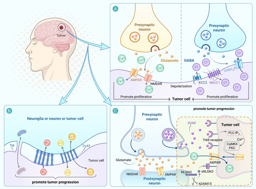
Figure 1.
Neural regulation of central nervous system tumors
-
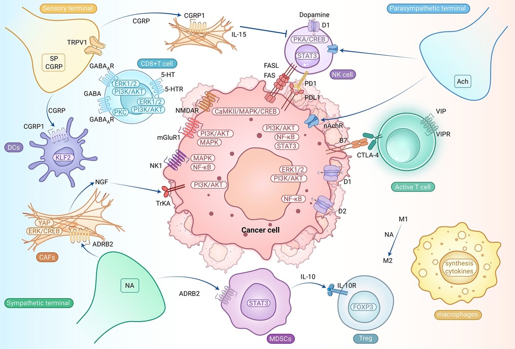
Figure 2.
Regulation of the TME by the peripheral nervous system
-
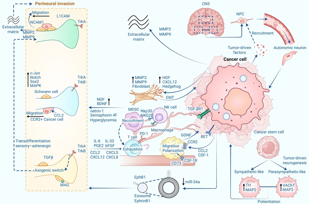
Figure 3.
Cancer cells hijack nerves, neuronal support cells, immune cells, and stromal cells to promote tumor progression
