
Multiview classification of pediatric medulloblastoma refines prognostic stratification and enables imaging-based risk prediction
-
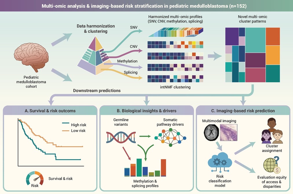
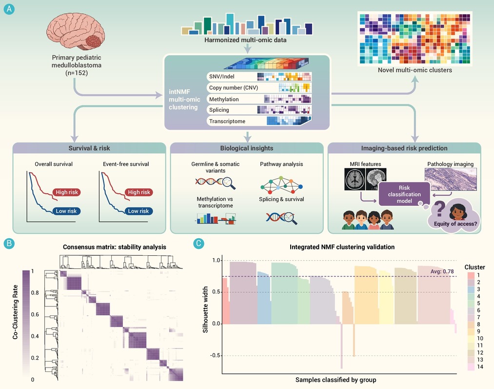
GRAPHICAL ABSTRACT
 DownLoad:
Full size image
DownLoad:
Full size image
-
PUBLIC SUMMARY
-
We developed novel multiomic classifications for pediatric medulloblastoma to improve risk stratification.
14 distinct molecular clusters reveal high- and low-risk populations that were previously grouped together.
Magnetic resonance and pathology images can predict molecular risk groups, improving accessibility.
This model offers a path toward reducing toxic drug effects and improving survival for aggressive disease.
-
-
ABSTRACT
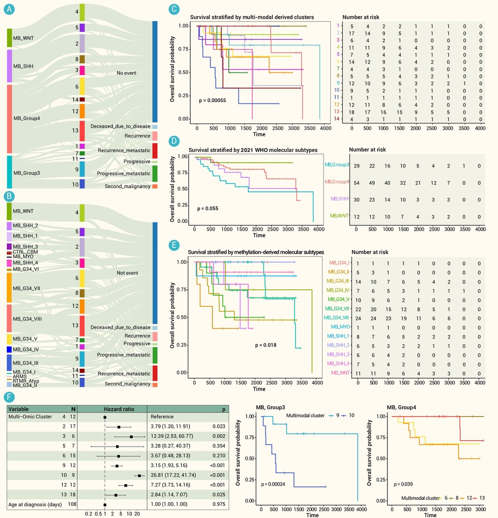
Despite advances in molecular subclassification, significant heterogeneity within the four primary medulloblastoma subgroups complicates therapeutic design. High-risk SHH and Group 3/4 cases, particularly those with metastases, frequently fail conventional therapy, highlighting the urgent need for molecularly-based risk refinement and improved disease estimation. By integrating five distinct molecular layers—non-synonymous variants, copy number variations (CNVs), DNA-methylation, gene expression, and alternative splicing—we identified 14 multi-omic clusters (MOCs) across 152 primary tumors from the Children’s Brain Tumor Network (CBTN). Within Group 3, we identified two prognostically distinct MOCs (p =0.00024 ); favorable outcomes were linked to high-fidelity genomic maintenance and proteostasis, while poor prognosis was characterized by Notch signaling and metabolic reprogramming (IDH2/3B). Similarly, Group 4 MOCs showed divergent outcomes (p = 0.039) driven by either RTK-mediated proliferation or epigenetic stability. Germline analysis revealed that chromatin-modifying variants (SIRT2, PRDM2) associated with favorable clusters, while invasion-associated variants (PTPRT, EPHA1) linked to tumor aggression. In an effort to explore potential avenues for clinical translation, we modeled MRI and histopathology features as surrogates for these molecularly-defined risks. This integrated multi-omic approach refines our understanding of medulloblastoma outcomes, suggesting that integrated omics coupled with clinical imaging can significantly enhance precision medicine and prognostication.-
Keywords:
- Risk Classification /
- Multiview Clustering
-

-
REFERENCES
[1] Northcott P.A., Buchhalter I., Morrissy A.S., et al. (2017). The whole-genome landscape of medulloblastoma subtypes. Nature 547:311−317. DOI:10.1038/nature22973 [2] Coltin H., Pequeno P., Liu N., et al. (2023). The Burden of surviving childhood medulloblastoma: A population-based, matched cohort study in Ontario, Canada. J. Clin. Oncol. 41:2372−2381. DOI:10.1200/JCO.22.02466 [3] Cavalli F.M., Remke M., Rampasek L., et al. (2017). Intertumoral heterogeneity within medulloblastoma subgroups. Cancer Cell 31:737−754.e736. DOI:10.1016/j.ccell.2017.05.005 [4] Louis D.N., Perry A., Wesseling P., et al. (2021). The 2021 WHO classification of tumors of the central nervous system: A summary. Neuro. Oncol. 23:1231−1251. DOI:10.1093/neuonc/noab106 [5] Sharma T., Schwalbe E.C., Williamson D., et al. (2019). Second-generation molecular subgrouping of medulloblastoma: An international meta-analysis of Group 3 and Group 4 subtypes. Acta Neuropathol. 138:309−326. DOI:10.1007/s00401-019-02020-0 [6] Smith K.S., Dhanda S.K., Billups C.A., et al. (2025). An integrated analysis of three medulloblastoma clinical trials refines risk-stratification approaches for reducing toxicity and improving survival. Neuro. Oncol. 28:268−281. DOI:10.1093/neuonc/noaf250 [7] Kumar R., Smith K.S., Deng M., et al. (2021). Clinical outcomes and patient-matched molecular composition of relapsed medulloblastoma. J. Clin. Oncol. 39:807−821. DOI:10.1200/JCO.20.01359 [8] Chalise P. and Fridley B.L. (2017). Integrative clustering of multi-level 'omic data based on non-negative matrix factorization algorithm. PLoS One 12:e0176278. DOI:10.1371/journal.pone.0176278 [9] Capper D., Jones D.T.W., Sill M., et al. (2018). DNA methylation-based classification of central nervous system tumours. Nature 555:469−474. DOI:10.1038/nature26000 [10] Silva T.C., Young J.I., Martin E.R., et al. (2022). MethReg: Estimating the regulatory potential of DNA methylation in gene transcription. Nucleic Acids Res. 50:e51. DOI:10.1093/nar/gkac030 [11] Juraschka K. and Taylor M.D. (2019). Medulloblastoma in the age of molecular subgroups: A review. J. Neurosurg. Pediatr. 24:353−363. DOI:10.3171/2019.5.PEDS18381 [12] Zou H., Poore B., Broniscer A., et al. (2020). Molecular heterogeneity and cellular diversity: Implications for precision treatment in medulloblastoma. Cancers (Basel) 12. DOI:10.3390/cancers12030643. [13] Tavtigian S.V., Harrison S.M., Boucher K.M., et al. (2020). Fitting a naturally scaled point system to the ACMG/AMP variant classification guidelines. Hum. Mutat. 41:1734−1737. DOI:10.1002/humu.24088 [14] Richards S., Aziz N., Bale S., et al. (2015). Standards and guidelines for the interpretation of sequence variants: A joint consensus recommendation of the American College of Medical Genetics and Genomics and the Association for Molecular Pathology. Genet. Med. 17:405−424. DOI:10.1038/gim.2015.30 [15] Northcott P.A., Korshunov A., Witt H., et al. (2011). Medulloblastoma comprises four distinct molecular variants. J. Clin. Oncol. 29:1408−1414. DOI:10.1200/JCO.2009.27.4324 [16] Northcott P.A., Shih D.J., Peacock J., et al. (2012). Subgroup-specific structural variation across 1,000 medulloblastoma genomes. Nature 488:49−56. DOI:10.1038/nature11327 [17] Lui V.W., Hedberg M.L., Li H., et al. (2013). Frequent mutation of the PI3K pathway in head and neck cancer defines predictive biomarkers. Cancer Discov. 3:761−769. DOI:10.1158/2159-8290.CD-13-0103 [18] Sikkema A.H., den Dunnen W.F., Hulleman E., et al. (2012). EphB2 activity plays a pivotal role in pediatric medulloblastoma cell adhesion and invasion. Neuro. Oncol. 14:1125−1135. DOI:10.1093/neuonc/nos130 [19] Jing H., Hu J., He B., et al. (2016). A SIRT2-Selective inhibitor promotes c-Myc oncoprotein degradation and exhibits broad anticancer activity. Cancer Cell 29:297−310. DOI:10.1016/j.ccell.2016.02.007 [20] Keenan A.B., Jenkins S.L., Jagodnik K.M., et al. (2018). The library of integrated network-based cellular signatures NIH program: System-level cataloging of human cells response to perturbations. Cell Syst. 6:13−24. DOI:10.1016/j.cels.2017.11.001 [21] Ouyang J., Zhang Y., Xiong F., et al. (2021). The role of alternative splicing in human cancer progression. Am. J. Cancer Res. 11:4642−4667. DOI:10.21236/ada477527 [22] Acevedo-Diaz A., Morales-Caban B.M., Zayas-Santiago A., et al. (2022). SCAMP3 regulates EGFR and promotes proliferation and migration of triple-negative breast cancer cells through the modulation of AKT, ERK, and STAT3 signaling pathways. Cancers (Basel) 14:2807. DOI:10.3390/cancers14112807 [23] Kumamoto T., Yamada K., Yoshida S., et al. (2020). Impairment of DYRK2 by DNMT1-mediated transcription augments carcinogenesis in human colorectal cancer. Int. J. Oncol. 56:1529−1539. DOI:10.3892/ijo.2020.5020 [24] Gad A.A. and Balenga N. (2020). The emerging role of adhesion GPCRs in cancer. ACS Pharmacol. Transl. Sci. 3:29−42. DOI:10.1021/acsptsci.9b00093 [25] Chase A., Ernst T., Fiebig A., et al. (2010). TFG, a target of chromosome translocations in lymphoma and soft tissue tumors, fuses to GPR128 in healthy individuals. Haematologica 95:20−26. DOI:10.3324/haematol.2009.011536 [26] Sawada G., Ueo H., Matsumura T., et al. (2013). CHD8 is an independent prognostic indicator that regulates Wnt/beta-catenin signaling and the cell cycle in gastric cancer. Oncol. Rep. 30:1137−1142. DOI:10.3892/or.2013.2597 [27] Zhang C., Ni X., Tao C., et al. (2024). Targeting PUF60 prevents tumor progression by retarding mRNA decay of oxidative phosphorylation in ovarian cancer. Cell. Oncol. (Dordr) 47:157−174. DOI:10.1007/s13402-023-00859-w [28] Wang J., Huang Z., Ji L., et al. (2022). SHF acts as a novel tumor suppressor in glioblastoma multiforme by disrupting STAT3 dimerization. Adv. Sci. (Weinh) 9:e2200169. DOI:10.1002/advs.202200169 [29] Pages M., Pajtler K.W., Puget S., et al. (2019). Diagnostics of pediatric supratentorial RELA ependymomas: Integration of information from histopathology, genetics, DNA methylation and imaging. Brain. Pathol. 29:325−335. DOI:10.1111/bpa.12664 [30] Liu L., Cui J., Zhao Y., et al. (2021). KDM6A-ARHGDIB axis blocks metastasis of bladder cancer by inhibiting Rac1. Mol. Cancer 20:77. DOI:10.1186/s12943-021-01369-9 [31] Chen L., Zhou J., Zhao Z., et al. (2022). Low expression of phosphodiesterase 2 (PDE2A) promotes the progression by regulating mitochondrial morphology and ATP content and predicts poor prognosis in hepatocellular carcinoma. Cells 12:68. DOI:10.3390/cells12010068 [32] Manousakis E., Miralles C.M., Esquerda M.G., et al. (2023). CDKN1A/p21 in breast cancer: Part of the problem, or part of the solution? Int. J. Mol. Sci. 24:17488. DOI:10.3390/ijms242417488 [33] Watanabe M., Nakahata S., Hamasaki M., et al. (2010). Downregulation of CDKN1A in adult T-cell leukemia/lymphoma despite overexpression of CDKN1A in human T-lymphotropic virus 1-infected cell lines. J. Virol. 84:6966−6977. DOI:10.1128/JVI.00073-10 [34] Yu J., Xie Y., Li M., et al. (2019). Association between SFRP promoter hypermethylation and different types of cancer: A systematic review and meta-analysis. Oncol. Lett. 18:3481−3492. DOI:10.3892/ol.2019.10709 [35] Chen Y.Z., Liu D., Zhao Y.X., et al. (2014). Aberrant promoter methylation of the SFRP1 gene may contribute to colorectal carcinogenesis: A meta-analysis. Tumour Biol. 35:9201−9210. DOI:10.1007/s13277-014-2180-x [36] Xie M., Wu X., Zhang J., et al. (2017). Ski regulates Smads and TAZ signaling to suppress lung cancer progression. Mol. Carcinog. 56:2178−2189. DOI:10.1002/mc.22661 [37] Deheuninck J. and Luo K. (2009). Ski and SnoN, potent negative regulators of TGF-beta signaling. Cell Res. 19:47−57. DOI:10.1038/cr.2008.324 [38] Xiong X., Yang C., Jin Y., et al. (2024). ABHD6 suppresses colorectal cancer progression via AKT signaling pathway. Mol. Carcinog. 63:647−662. DOI:10.1002/mc.23678 [39] Nitzki F., Tolosa E.J., Cuvelier N., et al. (2015). Overexpression of mutant Ptch in rhabdomyosarcomas is associated with promoter hypomethylation and increased Gli1 and H3K4me3 occupancy. Oncotarget 6:9113−9124. DOI:10.18632/oncotarget.3272 [40] Xu H., Huang K., Lin Y., et al. (2023). Glycosyltransferase GLT8D1 and GLT8D2 serve as potential prognostic biomarkers correlated with tumor immunity in gastric cancer. BMC Med. Genomics 16:123. DOI:10.1186/s12920-023-01559-y [41] Schwartzman J., Mongoue-Tchokote S., Gibbs A., et al. (2011). A DNA methylation microarray-based study identifies ERG as a gene commonly methylated in prostate cancer. Epigenetics 6:1248−1256. DOI:10.4161/epi.6.10.17727 [42] Wang S., Ding Y.B., Chen G.Y., et al. (2004). Hypermethylation of Syk gene in promoter region associated with oncogenesis and metastasis of gastric carcinoma. World J. Gastroenterol. 10:1815−1818. DOI:10.3748/wjg.v10.i12.1815 [43] Kunze E., Wendt M. and Schlott T. (2006). Promoter hypermethylation of the 14-3-3 σ, SYK and CAGE-1 genes is related to the various phenotypes of urinary bladder carcinomas and associated with progression of transitional cell carcinomas. Int. J. Mol. Med. 18:547−557. DOI:10.3892/ijmm.18.4.547 [44] Huang S., Wang C., Yi Y., et al. (2015). Kruppel-like factor 9 inhibits glioma cell proliferation and tumorigenicity via downregulation of miR-21. Cancer Lett. 356:547−555. DOI:10.1016/j.canlet.2014.10.007 [45] Pham K., Maxwell M.J., Sweeney H., et al. (2021). Novel glutamine antagonist JHU395 suppresses MYC-driven medulloblastoma growth and induces apoptosis. J. Neuropathol. Exp. Neurol. 80:336−344. DOI:10.1093/jnen/nlab018 [46] Takebe N., Miele L., Harris P.J., et al. (2015). Targeting Notch, Hedgehog, and Wnt pathways in cancer stem cells: Clinical update. Nat. Rev. Clin. Oncol. 12:445−464. DOI:10.1038/nrclinonc.2015.61 [47] Gwynne W.D., Suk Y., Custers S., et al. (2022). Cancer-selective metabolic vulnerabilities in MYC-amplified medulloblastoma. Cancer Cell 40:1488−1502.e1487. DOI:10.1016/j.ccell.2022.10.009 [48] Karaulic A., Fournier C. and Pages G. (2025). Exploring novel applications: Repositioning clinically approved therapies for medulloblastoma treatment. Cancers (Basel) 17. DOI:10.3390/cancers17223659 [49] Xiao H., Bid H.K., Jou D., et al. (2015). A novel small molecular STAT3 inhibitor, LY5, inhibits cell viability, cell migration, and angiogenesis in medulloblastoma cells. J. Biol. Chem. 290:3418−3429. DOI:10.1074/jbc.M114.616748 -
ABOUT THIS ARTICLE
Cite this article:
Kraya A., Rathi K., Kazerooni A. F., et al. (2026). Multiview classification of pediatric medulloblastoma refines prognostic stratification and enables imaging-based risk prediction. The Innovation Oncology 1:100025. https://doi.org/10.59717/j.xinn-oncol.2026.100025
Welcome!
To request copyright permission to republish or share portions of our works, please visit Copyright Clearance Center's (CCC) Marketplace website at marketplace.copyright.com.
Export File
Citation
Kraya A., Rathi K., Kazerooni A. F., et al. (2026). Multiview classification of pediatric medulloblastoma refines prognostic stratification and enables imaging-based risk prediction. The Innovation Oncology 1:100025. https://doi.org/10.59717/j.xinn-oncol.2026.100025

Format
Content
-
Figure 1.
Multi-omic clustering strategy and quality assessment of multi-omic clusters (MOCs)
-
Figure 2.
The relationship of MOCs with known subtypes and clinical outcomes
-
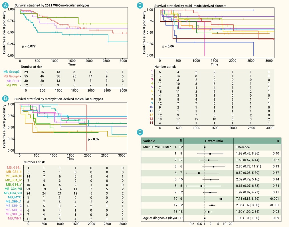
Figure 3.
Assessment of event-free survival (EFS) characteristics on the basis of four-subtype, second-generation subtype, and MOC classifications
-
Figure 4.
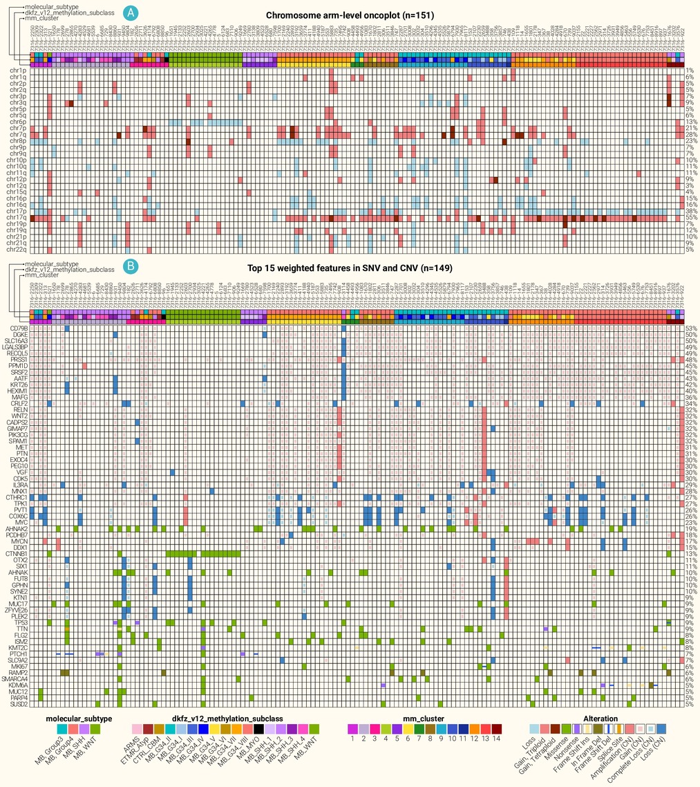
Somatic variation across medulloblastoma MOCs
-
Figure 5.
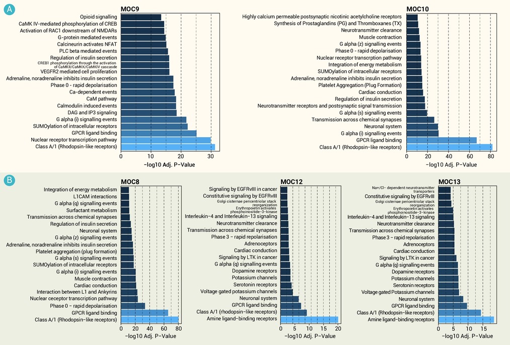
Cluster-specific transcriptomic characteristics across medulloblastoma MOCs
-
Figure 6.
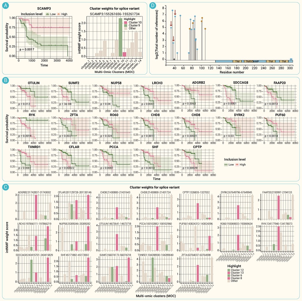
Prognostic differential splicing events identified across prognostically significant MOCs
-
Figure 7.
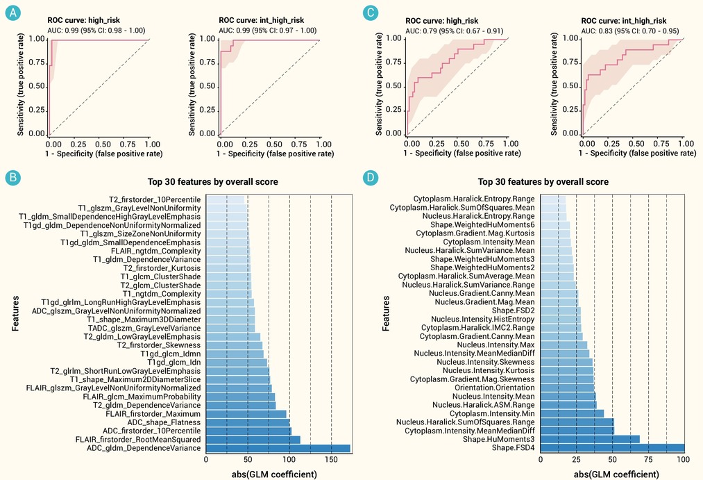
Elastic net logistic regression of MOC-based risk as a function of pre-operative MRI and pathomic features
