
Wnt signaling arranges the fate of lipid metabolism: The role of gut microbes
-
GRAPHICAL ABSTRACT
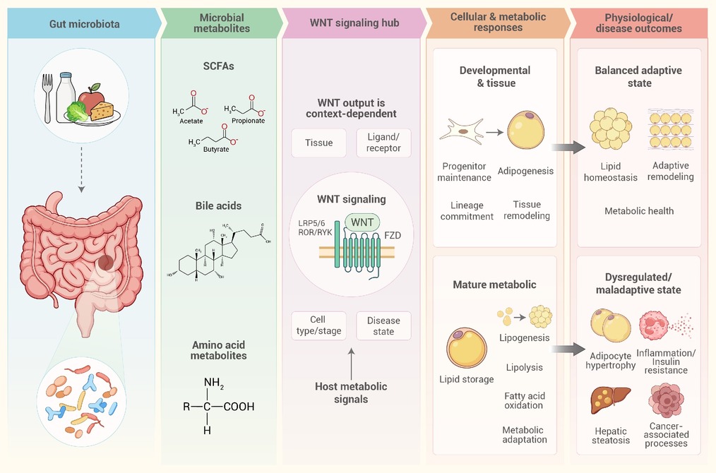
 DownLoad:
Full size image
-
PUBLIC SUMMARY
-
Wnt signaling has dual roles in fat-cell development and metabolism.
Metabolites from gut microbes can shift this balance, linking microbes to fat metabolism and tissue health.
Gut microbes, their metabolites, and Wnt signaling offer potential therapeutic targets.
-
-
ABSTRACT
Disrupted lipid metabolism contributes to metabolic diseases and cancer. Gut microbes and their metabolites influence lipid absorption, synthesis, storage, and oxidation, while the Wnt signaling pathway is not only associated with lipid metabolism as a downstream metabolic pathway, but also serves as a regulatory system linking adipocyte lineage determination, tissue remodeling, and specific metabolic functions in mature adipocytes. How these components interact to form a gut microbes–metabolites–Wnt signaling axis, however, remains incompletely understood. Thus, this review focuses on the structure and physiological functions of Wnt, the association between Wnt signaling and host lipid metabolism. This review then examines how bile acids, amino acid metabolites, and short-chain fatty acids intersect with Wnt-related pathways across intestinal, hepatic, adipose, and cancer models. It further discusses the targeting of the gut microbes–metabolites–Wnt signaling axis for treatment, and explored the application prospects and challenges of gut microbes and their metabolites in the prevention and treatment of metabolic diseases and cancer.-
Keywords:
- Wnt signaling /
- gut microbiota /
- lipid metabolism /
- metabolic diseases
-

-
REFERENCES
[1] Zarghamravanbakhsh P., Frenkel M. and Poretsky L. (2021). Metabolic causes and consequences of nonalcoholic fatty liver disease (NAFLD). Metab. Open. 12:100149. DOI:10.1016/j.metop.2021.100149 [2] Ma Y.F., Liu J.X. and Wang H.F. (2023). Porcine gut microbiota and lipid metabolism: Recent advances and future directions. Sci. Sin. Vitae. 53:616−625. DOI:10.1360/SSV-2021-0363 [3] Ross S.E., Hemati N., Longo K.A. et al. (2000). Inhibition of adipogenesis by Wnt signaling. Science 289:950−953. DOI:10.1126/science.289.5481.950 [4] Yang Loureiro Z., Joyce S., DeSouza T. et al. (2023). Wnt signaling preserves progenitor cell multipotency during adipose tissue development. Nat. Metab. 5:1014−1028. DOI:10.1038/s42255-023-00813-y [5] Bennett C.N., Ross S.E., Longo K.A. et al. (2002). Regulation of Wnt signaling during adipogenesis. J. Biol. Chem. 277:30998−31004. DOI:10.1074/jbc.M204527200 [6] Liu H., Li P., Zhang S. et al. (2022a). Prrx1 marks stem cells for bone, white adipose tissue and dermis in adult mice. Nat. Genet. 54:1946−1958. DOI:10.1038/s41588-022-01227-4 [7] Zeve D., Seo J., Suh J.M. et al. (2012). Wnt signaling activation in adipose progenitors promotes insulin-independent muscle glucose uptake. Cell Metab. 15:492−504. DOI:10.1016/j.cmet.2012.03.010 [8] Bagchi D.P. and MacDougald O.A. (2021). Wnt signaling: From mesenchymal cell fate to lipogenesis and other mature adipocyte functions. Diabetes 70:1419−1430. DOI:10.2337/dbi20-0015 [9] Liu S. (2025). Mechanisms of gut microbiota in host fat deposition: Metabolites, signaling pathways, and translational applications. Front. Microbiol. 16:1675155. DOI:10.3389/fmicb.2025.1675155 [10] Hemba-Waduge R.U.S., Liu M. and Ji J.Y. (2025). Transcriptional regulation of lipid metabolism by Wnt signaling and hox protein cues. Deng W.M. and González C. (eds). The Drosophila Model in Cancer 61:81. DOI:10.1007/978-3-031-97035-1_4 [11] Sanam M., Hossain C.F.T.Z., Hyder T.B. et al. (2025). Bridging two worlds: Host microbiota crosstalk in health and dysregulation. Innate Immun. 31:17534259251392993. DOI:10.1177/17534259251392993. [12] Willert K. and Nusse R. (2012). Wnt proteins. Cold Spring Harb. Perspect. Biol. 4:a007864−a007864. DOI:10.1101/cshperspect.a007864 [13] Yu J. and Virshup D.M. (2022). Functional regulation of Wnt protein through post-translational modifications. Biochem. Soc. Trans. 50:1797−1808. DOI:10.1042/BST20220735 [14] Janda C.Y., Waghray D., Levin A.M. et al. (2012). Structural basis of Wnt recognition by frizzled. Science 337:59−64. DOI:10.1126/science.1222879 [15] MacDonald B.T., Hien A., Zhang X. et al. (2014). Disulfide bond requirements for active Wnt ligands. J. Biol. Chem. 289:18122−18136. DOI:10.1074/jbc.M114.575027 [16] Liu Y., Qi X., Donnelly L. et al. (2022b). Mechanisms and inhibition of porcupine-mediated Wnt acylation. Nature 607:816−822. DOI:10.1038/s41586-022-04952-2 [17] Kurayoshi M., Yamamoto H., Izumi S. et al. (2007). Post-translational palmitoylation and glycosylation of Wnt-5a are necessary for its signalling. Biochem. J. 402:515−523. DOI:10.1042/BJ20061476 [18] Nygaard R., Yu J., Kim J. et al. (2021). Structural basis of WLS/Evi-mediated Wnt transport and secretion. Cell 184:194−206.e14. DOI:10.1016/j.cell.2020.11.038 [19] Zhong Q., Zhao Y., Ye F. et al. (2021). Cryo-EM structure of human Wntless in complex with Wnt3a. Nat. Commun. 12:4541. DOI:10.1038/s41467-021-24731-3 [20] Mikels A.J. and Nusse R. (2006). Purified Wnt5a protein activates or inhibits β-catenin–TCF signaling depending on receptor context. PLoS Biol. 4:e115. DOI:10.1371/journal.pbio.0040115 [21] Nusse R. and Clevers H. (2017). Wnt/β-catenin signaling, disease, and emerging therapeutic modalities. Cell 169:985−999. DOI:10.1016/j.cell.2017.05.016 [22] Qi X., Hu Q., Elghobashi-Meinhardt N. et al. (2023). Molecular basis of Wnt biogenesis, secretion, and Wnt7-specific signaling. Cell 186:5028−5040.e14. DOI:10.1016/j.cell.2023.09.021 [23] Miranda M., Galli L.M., Enriquez M. et al. (2014). Identification of the WNT1 residues required for palmitoylation by porcupine. FEBS Lett. 588:4815−4824. DOI:10.1016/j.febslet.2014.11.016 [24] Thomas K.R. and Capecchi M.R. (1990). Targeted disruption of the murine int-1 proto-oncogene resulting in severe abnormalities in midbrain and cerebellar development. Nature 346:847−850. DOI:10.1038/346847a0 [25] Laine C.M., Joeng K.S., Campeau P.M. et al. (2013). WNT1 mutations in early-onset osteoporosis and osteogenesis imperfecta. N. Engl. J. Med. 368:1809−1816. DOI:10.1056/NEJMoa1215458 [26] Mihara E., Hirai H., Yamamoto H. et al. (2016). Active and water-soluble form of lipidated Wnt protein is maintained by a serum glycoprotein afamin/α-albumin. eLife. 5:e11621. DOI:10.7554/eLife.11621 [27] Gao X. and Hannoush R.N. (2014). Single-cell imaging of Wnt palmitoylation by the acyltransferase porcupine. Nat. Chem. Biol. 10:61−68. DOI:10.1038/nchembio.1392 [28] Rios-Esteves J., Haugen B. and Resh M.D. (2014). Identification of key residues and regions important for porcupine-mediated Wnt acylation. J. Biol. Chem. 289:17009−17019. DOI:10.1074/jbc.M114.561209 [29] Takada S., Stark K.L., Shea M.J. et al. (1994). Wnt-3a regulates somite and tailbud formation in the mouse embryo. Genes Dev. 8:174−189. DOI:10.1101/gad.8.2.174 [30] Willert K., Brown J.D., Danenberg E. et al. (2003). Wnt proteins are lipid-modified and can act as stem cell growth factors. Nature 423:448−452. DOI:10.1038/nature01611 [31] de Winter T.J.J. and Nusse R. (2021). Running against the Wnt: How Wnt/β-catenin suppresses adipogenesis. Front. Cell Dev. Biol. 9:627429. DOI:10.3389/fcell.2021.627429 [32] Bauer M., Bénard J., Gaasterland T. et al. (2013). WNT5A encodes two isoforms with distinct functions in cancers. PLoS One. 8:e80526. DOI:10.1371/journal.pone.0080526 [33] Kamizaki K., Minami Y. and Nishita M. (2024). Role of the ror family receptors in Wnt5a signaling. In Vitro Cell. Dev. Biol. Anim. 60:489−501. DOI:10.1007/s11626-024-00885-4 [34] Hei Y., Hu Q., Manisa B. et al. (2024). Synthesis and frizzled-receptor binding of a WNT5A hairpin-3 peptide. Chem. Commun. 60:13534−13537. DOI:10.1039/D4CC04393F [35] Ge Y., de Almeida Magalhaes T., Wu H. et al. (2026). Conformational cycling of the Wntless transporter drives trafficking and secretion of Wnt morphogens. Nat. Commun. DOI:10.1038/s41467-026-74521-y. [36] Oishi I., Suzuki H., Onishi N. et al. (2003). The receptor tyrosine kinase Ror2 is involved in non-canonical Wnt5a/JNK signalling pathway. Genes Cells. 8:645−654. DOI:10.1046/j.1365-2443.2003.00662.x [37] McQuate A., Latorre-Esteves E. and Barria A. (2017). A Wnt/Calcium signaling cascade regulates Neuronal Excitability and Trafficking of NMDARs. Cell Rep. 21:60−69. DOI:10.1016/j.celrep.2017.09.023 [38] Yamaguchi T.P., Bradley A., McMahon A.P. et al. (1999). A Wnt5a pathway underlies outgrowth of multiple structures in the vertebrate embryo. Development 126:1211−1223. DOI:10.1242/dev.126.6.1211 [39] Ouchi N., Higuchi A., Ohashi K. et al. (2010). Sfrp5 is an anti-inflammatory adipokine that modulates metabolic dysfunction in obesity. Science 329:454−457. DOI:10.1126/science.1188280 [40] Fuster J.J., Zuriaga M.A., Ngo D.T.M. et al. (2015). Noncanonical Wnt signaling promotes obesity-induced adipose tissue inflammation and metabolic dysfunction independent of adipose tissue expansion. Diabetes 64:1235−1248. DOI:10.2337/db14-1164 [41] Zuriaga M.A., Fuster J.J., Farb M.G. et al. (2017). Activation of non-canonical WNT signaling in human visceral adipose tissue contributes to local and systemic inflammation. Sci. Rep. 7:17326. DOI:10.1038/s41598-017-17509-5 [42] Reggio A., Rosina M., Palma A. et al. (2020). Adipogenesis of skeletal muscle fibro/adipogenic progenitors is affected by the WNT5a/GSK3/β-catenin axis. Cell Death Differ. 27:2921−2941. DOI:10.1038/s41418-020-0551-y [43] Kirikoshi H., Sekihara H. and Katoh M. (2001a). Molecular cloning and characterization of human WNT7B. Int. J. Oncol. 19:779−783. DOI:10.3892/ijo.19.4.779 [44] Schmidt M., Poser C., Janster C. et al. (2022). The hairpin region of WNT7A is sufficient for binding to the Frizzled7 receptor and to elicit signaling in myogenic cells. Comput. Struct. Biotechnol. J. 20:6348−6359. DOI:10.1016/j.csbj.2022.10.047 [45] Cho C., Smallwood P.M. and Nathans J. (2017). Reck and Gpr124 are essential receptor cofactors for Wnt7a/Wnt7b-specific signaling in mammalian CNS angiogenesis and blood-brain barrier regulation. Neuron 95:1056−1073.e5. DOI:10.1016/j.neuron.2017.07.031 [46] Eubelen M., Bostaille N., Cabochette P. et al. (2018). A molecular mechanism for Wnt ligand-specific signaling. Science 361:eaat1178. DOI:10.1126/science.aat1178 [47] Le Grand F., Jones A.E., Seale V. et al. (2009). Wnt7a activates the planar cell polarity pathway to drive the symmetric expansion of satellite stem cells. Cell Stem Cell. 4:535−547. DOI:10.1016/j.stem.2009.03.013 [48] Parr B.A. and McMahon A.P. (1995). Dorsalizing signal Wnt-7a required for normal polarity of D–V and A–P axes of mouse limb. Nature 374:350−353. DOI:10.1038/374350a0 [49] Stenman J.M., Rajagopal J., Carroll T.J. et al. (2008). Canonical Wnt signaling regulates organ-specific assembly and differentiation of CNS vasculature. Science 322:1247−1250. DOI:10.1126/science.1164594 [50] Daneman R., Agalliu D., Zhou L. et al. (2009). Wnt/β-catenin signaling is required for CNS, but not non-CNS, angiogenesis. Proc. Natl. Acad. Sci. U.S.A. 106:641−646. DOI:10.1073/pnas.0805165106 [51] Shu W., Jiang Y.Q., Lu M.M. et al. (2002). Wnt7b regulates mesenchymal proliferation and vascular development in the lung. Development 129:4831−4842. DOI:10.1242/dev.129.20.4831 [52] Kirikoshi H., Sekihara H. and Katoh M. (2001b). WNT10A and WNT6, clustered in human chromosome 2q35 Region with head-to-tail manner, are strongly coexpressed in SW480 cells. Biochem. Biophys. Res. Commun. 283:798−805. DOI:10.1006/bbrc.2001.4855 [53] Benard E.L. and Hammerschmidt M. (2025). The fundamentals of WNT10A. Differentiation 142:100838. DOI:10.1016/j.diff.2025.100838 [54] Ain Q.U., Seemab U., Rashid S. et al. (2013). Prediction of structure of human WNT-CRD (FZD) complex for computational drug repurposing. PLoS One. 8:e54630. DOI:10.1371/journal.pone.0054630 [55] Hirai H., Matoba K., Mihara E. et al. (2019). Crystal structure of a mammalian Wnt–frizzled complex. Nat. Struct. Mol. Biol. 26:372−379. DOI:10.1038/s41594-019-0216-z [56] Cawthorn W.P., Bree A.J., Yao Y. et al. (2012). Wnt6, Wnt10a and Wnt10b inhibit adipogenesis and stimulate osteoblastogenesis through a β-catenin-dependent mechanism. Bone 50:477−489. DOI:10.1016/j.bone.2011.08.010 [57] Kang S., Bennett C. N., Gerin I. et al. (2007). Wnt signaling stimulates osteoblastogenesis of mesenchymal precursors by suppressing C/EBPα and PPARγ. J. Biol. Chem. 282:14515−14524. DOI:10.1074/jbc.M700030200 [58] Fang Q., Ye L., Han L. et al. (2025). LGR4 is a key regulator of hepatic gluconeogenesis. Free Radic. Biol. Med. 229:183−194. DOI:10.1016/j.freeradbiomed.2025.01.025 [59] Uno K., Uchino T., Suzuki T. et al. (2025). Rspo3-mediated metabolic liver zonation regulates systemic glucose metabolism and body mass in mice. PLoS Biol. 23:e3002955. DOI:10.1371/journal.pbio.3002955 [60] Ayala I., Hebbale S.K., Mononen J. et al. (2025). The spatial transcriptional activity of hepatic TCF7L2 regulates zonated metabolic pathways that contribute to liver fibrosis. Nat. Commun. 16:3408. DOI:10.1038/s41467-025-58714-5 [61] Silvano S., Napolitano T., Plaisant M. et al. (2025). RSPO1, a potent inducer of pancreatic β cell neogenesis. Cell Rep. Med. 6:102126. DOI:10.1016/j.xcrm.2025.102126 [62] Zhou A.X., Mondal T., Tabish A.M. et al. (2021). The long noncoding RNA TUNAR modulates Wnt signaling and regulates human β-cell proliferation. Am. J. Physiol. Endocrinol. Metab. 320:E846−E857. DOI:10.1152/ajpendo.00335.2020 [63] Kang S.W.S., Cunningham R.P., Miller C.B. et al. (2024). A spatial map of hepatic mitochondria uncovers functional heterogeneity shaped by nutrient-sensing signaling. Nat. Commun. 15:1799. DOI:10.1038/s41467-024-45751-9 [64] Badakhshi Y., Shao W., Liu D. et al. (2021). Estrogen-Wnt signaling cascade regulates expression of hepatic fibroblast growth factor 21. Am. J. Physiol. Endocrinol. Metab. 321:E292−E304. DOI:10.1152/ajpendo.00638.2020 [65] Song P., Gao Z., Bao Y. et al. (2024). Wnt/β-catenin signaling pathway in carcinogenesis and cancer therapy. J. Hematol. Oncol. 17:46. DOI:10.1186/s13045-024-01563-4 [66] Balatskyi V.V., Sowka A., Dobrzyn P. et al. (2023). WNT/β-catenin pathway is a key regulator of cardiac function and energetic metabolism. Acta Physiol. 237:e13912. DOI:10.1111/apha.13912 [67] Xu G., Chen T., Zhang Q. et al. (2026). Wnt/β-catenin signaling restrains developmental beige adipocyte thermogenesis and its inhibition imprints long-term energy expenditure. bioRxiv. DOI:10.64898/2026.03.23.713637. [68] Liu M., Hemba-Waduge R.U.S., Li X. et al. (2024). Wnt/Wingless signaling promotes lipid mobilization through signal-induced transcriptional repression. Proc. Natl. Acad. Sci. U.S.A. 121:e2322066121. DOI:10.1073/pnas.2322066121 [69] Yu F., Yu C., Li F. et al. (2021). Wnt/β-catenin signaling in cancers and targeted therapies. Signal Transduct Target Ther. 6:307. DOI:10.1038/s41392-021-00701-5 [70] Valvezan A.J., Zhang F., Diehl J.A. et al. (2012). Adenomatous polyposis coli (APC) regulates multiple signaling pathways by enhancing glycogen synthase kinase-3 (GSK-3) activity. J. Biol. Chem. 287:3823−3832. DOI:10.1074/jbc.M111.323337 [71] Niehrs C. and Acebron S.P. (2012). Mitotic and mitogenic Wnt signalling. EMBO J. 31:2705−2713. DOI:10.1038/emboj.2012.124 [72] Jeong W. and Jho E.H. (2021). Regulation of the low-density lipoprotein receptor-related protein LRP6 and its association with disease: Wnt/β-catenin signaling and beyond. Front. Cell Dev. Biol. 9:714330. DOI:10.3389/fcell.2021.714330 [73] González-Sancho J.M., Brennan K.R., Castelo-Soccio L.A. et al. (2004). Wnt proteins induce dishevelled phosphorylation via an LRP5/6- independent mechanism, irrespective of their ability to stabilize β-Catenin. Mol. Cell. Biol. 24:4757−4768. DOI:10.1128/MCB.24.11.4757-4768.2004 [74] Niehrs C. and Shen J. (2010). Regulation of Lrp6 phosphorylation. Cell. Mol. Life Sci. 67:2551−2562. DOI:10.1007/s00018-010-0329-3 [75] Krieghoff E., Behrens J.R. and Mayr B. (2006). Nucleo-cytoplasmic distribution of β-catenin is regulated by retention. J. Cell Sci. 119:1453−1463. DOI:10.1242/jcs.02864 [76] Lu F.I., Sun Y.H., Wei C.Y. et al. (2014). Tissue-specific derepression of TCF/LEF controls the activity of the Wnt/β-catenin pathway. Nat. Commun. 5:5368. DOI:10.1038/ncomms6368 [77] Shang S., Hua F. and Hu Z.W. (2017). The regulation of β-catenin activity and function in cancer: Therapeutic opportunities. Oncotarget 8:33972−33989. DOI:10.18632/oncotarget.15687 [78] Saito-Diaz K., Benchabane H., Tiwari A. et al. (2018). APC inhibits ligand-independent Wnt signaling by the clathrin endocytic pathway. Dev. Cell. 44:566−581.e8. DOI:10.1016/j.devcel.2018.02.013 [79] Chen L.J., Lin X.X., Guo J. et al. (2021). Lrp6 genotype affects individual susceptibility to nonalcoholic fatty liver disease and silibinin therapeutic response via Wnt/β-catenin-Cyp2e1 signaling. Int. J. Biol. Sci. 17:3936−3953. DOI:10.7150/ijbs.63732 [80] Bagchi D.P., Nishii A., Li Z. et al. (2020a). Wnt/β-catenin signaling regulates adipose tissue lipogenesis and adipocyte-specific loss is rigorously defended by neighboring stromal-vascular cells. Mol. Metab. 42:101078. DOI:10.1016/j.molmet.2020.101078 [81] Lefterova M.I., Zhang Y., Steger D.J. et al. (2008). PPARγ and C/EBP factors orchestrate adipocyte biology via adjacent binding on a genome-wide scale. Genes Dev. 22:2941−2952. DOI:10.1101/gad.1709008 [82] Mikkelsen T.S., Xu Z., Zhang X. et al. (2010). Comparative epigenomic analysis of murine and human adipogenesis. Cell 143:156−169. DOI:10.1016/j.cell.2010.09.006 [83] Bagchi D.P., Li Z., Corsa C.A. et al. (2020b). Wntless regulates lipogenic gene expression in adipocytes and protects against diet-induced metabolic dysfunction. Mol. Metab. 39:100992. DOI:10.1016/j.molmet.2020.100992 [84] Geoghegan G., Simcox J., Seldin M.M. et al. (2019). Targeted deletion of Tcf7l2 in adipocytes promotes adipocyte hypertrophy and impaired glucose metabolism. Mol. Metab. 24:44−63. DOI:10.1016/j.molmet.2019.03.003 [85] Akoumianakis I., Sanna F., Margaritis M. et al. (2019). Adipose tissue–derived WNT5A regulates vascular redox signaling in obesity via USP17/RAC1-mediated activation of NADPH oxidases. Sci. Transl. Med. 11:eaav5055. DOI:10.1126/scitranslmed.aav5055 [86] Bryja V., Andersson E.R., Schambony A. et al. (2009). The extracellular domain of Lrp5/6 inhibits noncanonical Wnt signaling in vivo. Mol. Biol. Cell. 20:924−936. DOI:10.1091/mbc.e08-07-0711 [87] van Amerongen R., Fuerer C., Mizutani M. et al. (2012). Wnt5a can both activate and repress Wnt/β-catenin signaling during mouse embryonic development. Dev. Biol. 369:101−114. DOI:10.1016/j.ydbio.2012.06.020 [88] Habas R., Kato Y. and He X. (2001). Wnt/Frizzled activation of rho regulates vertebrate gastrulation and requires a novel formin homology protein daam1. Cell. 107:843−854. DOI:10.1016/S0092-8674(01)00614-6 [89] Habas R., Dawid I.B. and He X. (2003). Coactivation of Rac and Rho by Wnt/Frizzled signaling is required for vertebrate gastrulation. Genes Dev. 17:295−309. DOI:10.1101/gad.1022203 [90] Bhatt P.M. and Malgor R. (2014). Wnt5a: A player in the pathogenesis of atherosclerosis and other inflammatory disorders. Atherosclerosis. 237:155−162. DOI:10.1016/j.atherosclerosis.2014.08.027 [91] Jing L., Wang H., Xia S. et al. (2025). Wnt/Ca2+ signaling: Dichotomous roles in regulating tumor progress (Review). Oncol. Lett. 30:1−12. DOI:10.3892/ol.2025.15145 [92] Slusarski D.C., Corces V.G. and Moon R.T. (1997). Interaction of Wnt and a Frizzled homologue triggers G-protein-linked phosphatidylinositol signalling. Nature 390:410−413. DOI:10.1038/37138 [93] Kohn A.D. and Moon R.T. (2005). Wnt and calcium signaling: β-catenin-independent pathways. Cell Calcium. 38:439−446. DOI:10.1016/j.ceca.2005.06.022 [94] Sheldahl L.C., Park M., Malbon C.C. et al. (1999). Protein kinase C is differentially stimulated by Wnt and Frizzled homologs in a G-protein-dependent manner. Curr. Biol. 9:695−S1. DOI:10.1016/S0960-9822(99)80310-8 [95] Kim Y.H. and Pyo S. (2019). Interleukin-10 suppresses adipogenesis via Wnt5a signaling pathway in 3T3-L1 preadipocytes. Biochem. Biophys. Res. Commun. 509:877−885. DOI:10.1016/j.bbrc.2019.01.033 [96] Bilkovski R., Schulte D.M., Oberhauser F. et al. (2010). Role of Wnt-5a in the determination of human mesenchymal stem cells into preadipocytes. J. Biol. Chem. 285:6170−6178. DOI:10.1074/jbc.M109.054338 [97] Kanazawa A., Tsukada S., Kamiyama M. et al. (2005). Wnt5b partially inhibits canonical Wnt/β-catenin signaling pathway and promotes adipogenesis in 3T3-L1 preadipocytes. Biochem. Biophys. Res. Commun. 330:505−510. DOI:10.1016/j.bbrc.2005.03.007 [98] van Tienen F.H.J., Laeremans H., van der Kallen C.J.H. et al. (2009). Wnt5b stimulates adipogenesis by activating PPARγ, and inhibiting the β-catenin dependent Wnt signaling pathway together with Wnt5a. Biochem. Biophys. Res. Commun. 387:207−211. DOI:10.1016/j.bbrc.2009.07.004 [99] Nishizuka M., Koyanagi A., Osada S. et al. (2008). Wnt4 and Wnt5a promote adipocyte differentiation. FEBS Lett. 582:3201−3205. DOI:10.1016/j.febslet.2008.08.011 [100] Menck K., Heinrichs S., Baden C. et al. (2021). The WNT/ROR pathway in cancer: From signaling to therapeutic intervention. Cell 10:142. DOI:10.3390/cells10010142 [101] Farb M.G., Karki S., Park S.Y. et al. (2016). WNT5A-JNK regulation of vascular insulin resistance in human obesity. Vasc. Med. 21:489−496. DOI:10.1177/1358863X16666693 [102] Tang Q., Chen C., Zhang Y. et al. (2018). Wnt5a regulates the cell proliferation and adipogenesis via MAPK-independent pathway in early stage of obesity. Cell Biol. Int. 42:63−74. DOI:10.1002/cbin.10862 [103] Zhu L., Shahein A., Choudhury S. et al. (2018). Upregulation of non-canonical Wnt ligands and oxidative glucose metabolism in NASH induced by methionine-choline-deficient diet. Trends Cell Mol. Biol. 13:47. DOI:10.31300/TCMB.13.2018.47-56 [104] Carbone C., Piro G., Gaianigo N. et al. (2018). Adipocytes sustain pancreatic cancer progression through a non-canonical WNT paracrine network inducing ROR2 nuclear shuttling. Int. J. Obes. 42:334−343. DOI:10.1038/ijo.2017.285 [105] Cabrae R., Dubuquoy C., Caüzac M. et al. (2020). Insulin activates hepatic Wnt/β-catenin signaling through stearoyl-CoA desaturase 1 and porcupine. Sci. Rep. 10:5186. DOI:10.1038/s41598-020-61869-4 [106] Guo F., Seldin M., Péterfy M. et al. (2021). NOTUM promotes thermogenic capacity and protects against diet-induced obesity in male mice. Sci. Rep. 11:16409. DOI:10.1038/s41598-021-95720-1 [107] Jiang H., Tao X., Yu S. et al. (2026). Adipocytic sclerostin loop3-LRP4 interaction required by sclerostin to impair whole-body lipid and glucose metabolism. Nat. Commun. 17:1812. DOI:10.1038/s41467-026-68526-w [108] Procházková N., Laursen M.F., La Barbera G. et al. (2024). Gut physiology and environment explain variations in human gut microbiome composition and metabolism. Nat. Microbiol. 9:3210−3225. DOI:10.1038/s41564-024-01856-x [109] Lin X., He K., Gu Z. et al. (2024). Emerging chemophysiological diversity of gut microbiota metabolites. Trends Pharmacol. Sci. 45:824−838. DOI:10.1016/j.tips.2024.07.006 [110] Cheng B., Wang Y.X., Wang Y.T. et al. (2025). Primary-secondary-tertiary axis of bile acid metabolism in healthy subjects. J. Proteome Res. 24:5572−5584. DOI:10.1021/acs.jproteome.5c00343 [111] Zhang J., Lyu A. and Wang C. (2023a). The molecular insights of bile acid homeostasis in host diseases. Life Sci. 330:121919. DOI:10.1016/j.lfs.2023.121919 [112] Fleishman J.S. and Kumar S. (2024). Bile acid metabolism and signaling in health and disease: Molecular mechanisms and therapeutic targets. Signal Transduct. Target. Ther. 9:97. DOI:10.1038/s41392-024-01811-6 [113] Ridlon J.M., Kang D. J., Hylemon P. B. (2006). Bile salt biotransformations by human intestinal bacteria. J. Lipid Res. 47:241−259. DOI:10.1194/jlr.R500013-JLR200 [114] Sayin S.I., Wahlström A., Felin J. et al. (2013). Gut microbiota regulates bile acid metabolism by reducing the levels of Tauro-beta-muricholic acid, a naturally occurring FXR antagonist. Cell Metab. 17:225−235. DOI:10.1016/j.cmet.2013.01.003 [115] Jia E.T., Liu Z.Y., Pan M. et al. (2019). Regulation of bile acid metabolism-related signaling pathways by gut microbiota in diseases. J. Zhejiang Univ. Sci. B. 20:781−792. DOI:10.1631/jzus.B1900073 [116] Gougelet A., Torre C., Veber P. et al. (2014). T-cell factor 4 and β-catenin chromatin occupancies pattern zonal liver metabolism in mice. Hepatology 59:2344−2357. DOI:10.1002/hep.26924 [117] Behari J., Yeh T.H., Krauland L. et al. (2010). Liver-specific β-catenin knockout mice exhibit defective bile acid and cholesterol homeostasis and increased susceptibility to diet-induced steatohepatitis. Am. J. Pathol. 176:744−753. DOI:10.2353/ajpath.2010.090667 [118] Yan W., Zhang K., Guo J. et al. (2025). Bile acid-mediated gut-liver axis crosstalk: The role of nuclear receptor signaling in dynamic regulation of inflammatory networks. Front. Immunol. 16:1595486. DOI:10.3389/fimmu.2025.1595486 [119] Thompson M.D., Moghe A., Cornuet P. et al. (2018). β-catenin regulation of farnesoid X receptor signaling and bile acid metabolism during murine cholestasis. Hepatology 67:955−971. DOI:10.1002/hep.29371 [120] Li J.Y., Gillilland M., Lee A.A. et al. (2022). Secondary bile acids mediate high-fat diet–induced upregulation of R-spondin 3 and intestinal epithelial proliferation. JCI Insight. 7:e148309. DOI:10.1172/jci.insight.148309 [121] Dong X., Cai C. and Fu T. (2023). FXR suppresses colorectal cancer by inhibiting the Wnt/β-catenin pathway via activation of TLE3. Genes Diss. 10:719−722. DOI:10.1016/j.gendis.2022.09.006 [122] Liu M., Sun J., Jia Z. et al. (2026). Medicago Sativa L. Saponin-driven lactobacillus intestinalis restores intestinal stemness in naturally aged mice via the bile acid-FXR-Wnt signaling axis. Adv. Sci. 13:e15370. DOI:10.1002/advs.202515370 [123] Holt J. A., Luo G., Billin A. N. et al. (2003). Definition of a novel growth factor-dependent signal cascade for the suppression of bile acid biosynthesis. Genes Dev. 17:1581−1591. DOI:10.1101/gad.1083503 [124] Inagaki T., Choi M., Moschetta A. et al. (2005). Fibroblast growth factor 15 functions as an enterohepatic signal to regulate bile acid homeostasis. Cell Metab. 2:217−225. DOI:10.1016/j.cmet.2005.09.001 [125] Zhang J. H., Nolan J. D., Kennie S. L. et al. (2013). Potent stimulation of fibroblast growth factor 19 expression in the human ileum by bile acids. Am. J. Physiol. Gastrointest. Liver Physiol. 304:G940−G948. DOI:10.1152/ajpgi.00398.2012 [126] Li X., Lu W., Kharitonenkov A. et al. (2024). Targeting the FGF19–FGFR4 pathway for cholestatic, metabolic, and cancerous diseases. J. Intern. Med. 295:292−312. DOI:10.1111/joim.13767 [127] Bhat N., Esteghamat F., Chaube B.K. et al. (2022). TCF7L2 transcriptionally regulates Fgf15 to maintain bile acid and lipid homeostasis through gut-liver crosstalk. FASEB J. 36:e22185. DOI:10.1096/fj.202101607R [128] Rinella M.E., Lieu H.D., Kowdley K.V. et al. (2024). A randomized, double-blind, placebo-controlled trial of aldafermin in patients with NASH and compensated cirrhosis. Hepatology 79:674−689. DOI:10.1097/HEP.0000000000000607 [129] Gao J., Xu K., Liu H. et al. (2018). Impact of the gut microbiota on intestinal immunity mediated by tryptophan metabolism. Front. Cell. Infect. Microbiol. 8:13. DOI:10.3389/fcimb.2018.00013 [130] Williams B.B., Van Benschoten A.H., Cimermancic P. et al. (2014). Discovery and characterization of gut microbiota decarboxylases that can produce the neurotransmitter tryptamine. Cell Host Microbe. 16:495−503. DOI:10.1016/j.chom.2014.09.001 [131] Dodd D., Spitzer M. H., Van Treuren W. et al. (2017). A gut bacterial pathway metabolizes aromatic amino acids into nine circulating metabolites. Nature 551:648−652. DOI:10.1038/nature24661 [132] Kurnasov O., Jablonski L., Polanuyer B. et al. (2003). Aerobic tryptophan degradation pathway in bacteria: Novel kynurenine formamidase. FEMS Microbiol. Lett. 227:219−227. DOI:10.1016/S0378-1097(03)00684-0 [133] Bortolotti P., Hennart B., Thieffry C. et al. (2016). Tryptophan catabolism in Pseudomonas aeruginosa and potential for inter-kingdom relationship. BMC Microbiol. 16:137. DOI:10.1186/s12866-016-0756-x [134] Zheng W., Liu M., Lv X. et al. (2025). AhR governs lipid metabolism: The role of gut microbiota. Front. Microbiol. 16:1442282. DOI:10.3389/fmicb.2025.1442282 [135] Metidji A., Omenetti S., Crotta S. et al. (2018). The environmental sensor AHR protects from inflammatory damage by maintaining intestinal stem cell homeostasis and barrier integrity. Immunity 49:353−362.e5. DOI:10.1016/j.immuni.2018.07.010 [136] Zhang L., Ji Q., Chen Q. et al. (2023b). Akkermansia muciniphila inhibits tryptophan metabolism via the AhR/β-catenin signaling pathway to counter the progression of colorectal cancer. Int. J. Biol. Sci. 19:4393−4410. DOI:10.7150/ijbs.85712 [137] Lee R., Li J., Li J. et al. (2022). Synthetic essentiality of tryptophan 2,3-dioxygenase 2 in APC -mutated colorectal cancer. Cancer Discov. 12:1702−1717. DOI:10.1158/2159-8290.CD-21-0680 [138] Huang T., Song J., Gao J. et al. (2022). Adipocyte-derived kynurenine promotes obesity and insulin resistance by activating the AhR/STAT3/IL-6 signaling. Nat. Commun. 13:3489. DOI:10.1038/s41467-022-31126-5 [139] Du M.X., Wang W., Jiang M.Z. et al. (2026). Christensenella massiliensis reduces kynurenine levels and alleviates obesity and related metabolic disorders in model mice. Gut Microbes. 18:2701382. DOI:10.1080/19490976.2026.2701382 [140] Nakagawa S., Yamaguchi K., Takane K. et al. (2024). Wnt/β-catenin signaling regulates amino acid metabolism through the suppression of CEBPA and FOXA1 in liver cancer cells. Commun. Biol. 7:510. DOI:10.1038/s42003-024-06202-9 [141] Madsen M. S., Siersbæk R., Boergesen M. et al. (2014). Peroxisome proliferator-activated receptor γ and C/EBPα synergistically activate key metabolic adipocyte genes by assisted loading. Mol. Cell. Biol. 34:939−954. DOI:10.1128/MCB.01344-13 [142] Pedersen H. K., Gudmundsdottir V., Nielsen H. B. et al. (2016). Human gut microbes impact host serum metabolome and insulin sensitivity. Nature 535:376−381. DOI:10.1038/nature18646 [143] Poncet N., Halley P.A., Lipina C. et al. (2020). Wnt regulates amino acid transporter Slc7a5 and so constrains the integrated stress response in mouse embryos. EMBO Rep. 21:EMBR201948469. DOI:10.15252/embr.201948469 [144] Wang D.Z., Chen G.Y., Li Y.F. et al. (2020). Comprehensive analysis of long non-coding RNA and mRNA expression profile in rectal cancer. Chin. Med. J. 133:1312−1321. DOI:10.1097/CM9.0000000000000753 [145] Wolfson R.L., Chantranupong L., Saxton R.A. et al. (2016). Sestrin2 is a leucine sensor for the mTORC1 pathway. Science 351:43−48. DOI:10.1126/science.aab2674 [146] Nicklin P., Bergman P., Zhang B. et al. (2009). Bidirectional transport of amino acids regulates mTOR and autophagy. Cell 136:521−534. DOI:10.1016/j.cell.2008.11.044 [147] Ricoult S.J.H. and Manning B.D. (2013). The multifaceted role of mTORC1 in the control of lipid metabolism. EMBO Rep. 14:242−251. DOI:10.1038/embor.2013.5 [148] Du D., Liu C., Qin M. et al. (2022). Metabolic dysregulation and emerging therapeutical targets for hepatocellular carcinoma. Acta Pharm. Sin. B. 12:558−580. DOI:10.1016/j.apsb.2021.09.019 [149] Tran T.Q., Hanse E.A., Habowski A.N. et al. (2020). α-Ketoglutarate attenuates Wnt signaling and drives differentiation in colorectal cancer. Nat. Cancer. 1:345−358. DOI:10.1038/s43018-020-0035-5 [150] Yochum G. S., Sherrick C. M., MacPartlin M. et al. (2010). A β-catenin/TCF-coordinated chromatin loop at MYC integrates 5′ and 3′ Wnt-responsive enhancers. Proc. Natl. Acad. Sci. U.S.A. 107:145−150. DOI:10.1073/pnas.0912294107 [151] Felsher D. W., and Bishop J.M. (1999). Reversible tumorigenesis by MYC in hematopoietic lineages. Mol. Cell. 4:199−207. DOI:10.1016/S1097-2765(00)80367-6 [152] Venkateswaran N., Lafita-Navarro M.C., Hao Y.H. et al. (2019). MYC promotes tryptophan uptake and metabolism by the kynurenine pathway in colon cancer. Genes Dev. 33:1236−1251. DOI:10.1101/gad.327056.119 [153] Zhao X., Petrashen A.P., Sanders J.A. et al. (2019). SLC1A5 glutamine transporter is a target of MYC and mediates reduced mTORC1 signaling and increased fatty acid oxidation in long-lived Myc hypomorphic mice. Aging Cell. 18:e12947. DOI:10.1111/acel.12947 [154] Wong C.C., Xu J., Bian X. et al. (2020). In colorectal cancer cells with mutant KRAS, SLC25A22-mediated glutaminolysis reduces DNA demethylation to increase WNT signaling, stemness, and drug resistance. Gastroenterology 159:2163−2180.e6. DOI:10.1053/j.gastro.2020.08.016 [155] Koh A., De Vadder F., Kovatcheva-Datchary P. et al. (2016). From dietary fiber to host physiology: Short-chain fatty acids as key bacterial metabolites. Cell 165:1332−1345. DOI:10.1016/j.cell.2016.05.041 [156] Louis P. and Flint H.J. (2017). Formation of propionate and butyrate by the human colonic microbiota. Environ. Microbiol. 19:29−41. DOI:10.1111/1462-2920.13589 [157] Bordonaro M., Lazarova D. L., and Sartorelli A. C. (2002). Cell type- and promoter-dependent modulation of the Wnt signaling pathway by sodium butyrate. Int. J. Cancer. 97:42−51. DOI:10.1002/ijc.1577 [158] Lazarova D.L., Chiaro C., Wong T. et al. (2013). CBP activity mediates effects of the histone deacetylase inhibitor butyrate on WNT activity and apoptosis in colon cancer cells. J. Cancer. 4:481−490. DOI:10.7150/jca.6583 [159] Chen J., Zhao K.N. and Vitetta L. (2019). Effects of intestinal microbial–elaborated butyrate on oncogenic signaling pathways. Nutrients 11:1026. DOI:10.3390/nu11051026 [160] Garavaglia B., Vallino L., Ferraresi A. et al. (2022). Butyrate inhibits colorectal cancer cell proliferation through autophagy degradation of β-catenin regardless of APC and β-catenin mutational status. Biomedicines 10:1131. DOI:10.3390/biomedicines10051131 [161] Chen D., Jin D., Huang S. et al. (2020). Clostridium butyricum, a butyrate-producing probiotic, inhibits intestinal tumor development through modulating Wnt signaling and gut microbiota. Cancer Lett. 469:456−467. DOI:10.1016/j.canlet.2019.11.019 [162] Ni M., Wang Z., Li Z. et al. (2025). Dietary supplement of sodium butyrate improves the growth performance and intestinal health by targeting Wnt/β-catenin signaling pathway in rabbits. Anim. Res. One Health. 3:389−404. DOI:10.1002/aro2.71 [163] Tyagi A.M., Yu M., Darby T.M. et al. (2018). The microbial metabolite butyrate stimulates bone formation via T regulatory cell-mediated regulation of WNT10B expression. Immunity 49:1116−1131.e7. DOI:10.1016/j.immuni.2018.10.013 [164] Tolhurst G., Heffron H., Lam Y.S. et al. (2012). Short-chain fatty acids stimulate glucagon-like peptide-1 secretion via the G-protein–coupled receptor FFAR2. Diabetes. 61:364−371. DOI:10.2337/db11-1019 [165] Geng H., Zhou W., Zhu B. et al. (2024). Intestinal flora modulates blood glucose via regulating the secretion of GLP-1 induced by propionate in mouse models of gestational diabetes mellitus. Research Square. DOI:10.21203/rs.3.rs-5276950/v1 [166] Chambers E.S., Viardot A., Psichas A. et al. (2015). Effects of targeted delivery of propionate to the human colon on appetite regulation, body weight maintenance and adiposity in overweight adults. Gut 64:1744−1754. DOI:10.1136/gutjnl-2014-307913 [167] den Besten G., Bleeker A., Gerding A. et al. (2015). Short-chain fatty acids protect against high-fat diet–induced obesity via a PPARγ-dependent switch from lipogenesis to fat oxidation. Diabetes 64:2398−2408. DOI:10.2337/db14-1213 [168] Kimura I., Ozawa K., Inoue D. et al. (2013). The gut microbiota suppresses insulin-mediated fat accumulation via the short-chain fatty acid receptor GPR43. Nat. Commun. 4:1829. DOI:10.1038/ncomms2852 [169] Gao Z., Yin J., Zhang J. et al. (2009). Butyrate improves insulin sensitivity and increases energy expenditure in mice. Diabetes 58:1509−1517. DOI:10.2337/db08-1637 [170] Yang J., Li G., Wang S. et al. (2025). Butyrate prevents obesity accompanied by HDAC9-mediated browning of white adipose tissue. Biomedicines 13:260. DOI:10.3390/biomedicines13020260 [171] Yang L., Huang Y., Chen M. et al. (2026). Pasteurized Akkermansia muciniphila AKK PROBIO attenuates obesity through gut Microbiota-SCFA-GLP-1 axis and potential involvement of AMPK/PPAR-α pathway. Probiotics Antimicrob. Proteins. 18:4812−4829. DOI:10.1007/s12602-025-10805-x [172] Yu H., Li R., Huang H. et al. (2018). Short-chain fatty acids enhance the lipid accumulation of 3T3-L1 cells by modulating the expression of enzymes of fatty acid metabolism. Lipids 53:77−84. DOI:10.1002/lipd.12005 [173] Jiao A., Yu B., He J. et al. (2020). Short chain fatty acids could prevent fat deposition in pigs via regulating related hormones and genes. Food Funct. 11:1845−1855. DOI:10.1039/C9FO02585E [174] Sun Y., Zhang J., Hong J. et al. (2023). Human RSPO1 mutation represses beige adipocyte thermogenesis and contributes to diet-induced adiposity. Adv. Sci. 10:2207152. DOI:10.1002/advs.202207152 [175] Gong J., Shen Y., Zhang H. et al. (2022). Gut microbiota characteristics of people with obesity by Meta-analysis of existing datasets. Nutrients 14:2993. DOI:10.3390/nu14142993 [176] Rodrigues V.F., Elias-Oliveira J., Pereira Í.S. et al. (2022). Akkermansia muciniphila and gut immune system: A good friendship that attenuates inflammatory bowel disease, obesity, and diabetes. Front. Immunol. 13:934695. DOI:10.3389/fimmu.2022.934695 [177] Ma X., Li M., Zhang Y. et al. (2025). Akkermansia muciniphila identified as key strain to alleviate gut barrier injury through Wnt signaling pathway. eLife 12:RP92906. DOI:10.7554/eLife.92906.5 [178] Badralmaa Y. and Natarajan V. (2025). Aberrant Wnt/β-catenin signaling in the mesenchymal stem cells of LZTFL1-depleted mice leads to increased adipogenesis, with implications for obesity. J. Biol. Chem. 301:108057. DOI:10.1016/j.jbc.2024.108057 [179] Emont M.P., Jacobs C., Essene A.L. et al. (2022). A single-cell atlas of human and mouse white adipose tissue. Nature 603:926−933. DOI:10.1038/s41586-022-04518-2 [180] Massier L., Jalkanen J., Elmastas M. et al. (2023). An integrated single cell and spatial transcriptomic map of human white adipose tissue. Nat. Commun. 14:1438. DOI:10.1038/s41467-023-36983-2 [181] Shao M., Vishvanath L., Busbuso N.C. et al. (2018). De novo adipocyte differentiation from Pdgfrβ+ preadipocytes protects against pathologic visceral adipose expansion in obesity. Nat. Commun. 9:890. DOI:10.1038/s41467-018-03196-x [182] Braud L., Bernabe M., Vernerey J. et al. (2025). TERT expression attenuates metabolic disorders in obese mice by promoting adipose stem and progenitor cell expansion and differentiation. Mol. Metab. 102:102262. DOI:10.1016/j.molmet.2025.102262 [183] Wang J., Liu R., Wang F. et al. (2013). Ablation of LGR4 promotes energy expenditure by driving white-to-brown fat switch. Nat. Cell Biol. 15:1455−1463. DOI:10.1038/ncb2867 [184] Zhang Z., Wang L., Qiao H. et al. (2025). Cryo-EM structure of the full-length LGR4-RSPOs complex and a targeting nanobody for anti-obesity therapy. Nat. Commun. 16:8406. DOI:10.1038/s41467-025-63410-5 [185] Dougherty M.W. and Jobin C. (2023). Intestinal bacteria and colorectal cancer: Etiology and treatment. Gut Microbes. 15:2185028. DOI:10.1080/19490976.2023.2185028 [186] Rubinstein M.R., Wang X., Liu W. et al. (2013). Fusobacterium nucleatum promotes colorectal carcinogenesis by modulating E-cadherin/β-catenin signaling via its FadA adhesin. Cell Host Microbe. 14:195−206. DOI:10.1016/j.chom.2013.07.012 [187] Dadgar-Zankbar L., Shariati A., Bostanghadiri N. et al. (2023). Evaluation of enterotoxigenic bacteroides fragilis correlation with the expression of cellular signaling pathway genes in Iranian patients with colorectal cancer. Infect. Agents Cancer. 18:48. DOI:10.1186/s13027-023-00523-w [188] Kang X., Ng S.K., Liu C. et al. (2023). Altered gut microbiota of obesity subjects promotes colorectal carcinogenesis in mice. eBioMedicine 93:104670. DOI:10.1016/j.ebiom.2023.104670 [189] Parida S., Wu S., Siddharth S. et al. (2021). A procarcinogenic colon microbe promotes breast tumorigenesis and metastatic progression and concomitantly activates notch and β-catenin axes. Cancer Discov. 11:1138−1157. DOI:10.1158/2159-8290.CD-20-0537 [190] Cui B., Luo H., He B. et al. (2025). Gut dysbiosis conveys psychological stress to activate LRP5/β-catenin pathway promoting cancer stemness. Signal Transduct. Target. Ther. 10:79. DOI:10.1038/s41392-025-02159-1 [191] Lv J., Jin S., Zhou Y. et al. (2025). Gut microbiota-derived metabolite phenylacetylglutamine inhibits the progression of prostate cancer by suppressing the Wnt/β-catenin signaling pathway. Front. Pharmacol. 16:1528058. DOI:10.3389/fphar.2025.1528058 [192] Agus A., Clément K. and Sokol H. (2021). Gut microbiota-derived metabolites as central regulators in metabolic disorders. Gut 70:1174−1182. DOI:10.1136/gutjnl-2020-323071 [193] Michael D. R., Jack A. A., Masetti G. et al. (2020). A randomised controlled study shows supplementation of overweight and obese adults with lactobacilli and bifidobacteria reduces bodyweight and improves well-being. Sci. Rep. 10:4183. DOI:10.1038/s41598-020-60991-7 [194] Depommier C., Everard A., Druart C. et al. (2019). Supplementation with Akkermansia muciniphila in overweight and obese human volunteers: a proof-of-concept exploratory study. Nat. Med. 25:1096−1103. DOI:10.1038/s41591-019-0495-2 [195] Chen Z., Guo L., Zhang Y. et al. (2014). Incorporation of therapeutically modified bacteria into gut microbiota inhibits obesity. J. Clin. Invest. 124:3391−3406. DOI:10.1172/JCI72517 [196] Tian Q., Zhao J., Yang Q. et al. (2020). Dietary alpha-ketoglutarate promotes beige adipogenesis and prevents obesity in middle-aged mice. Aging Cell. 19:e13059. DOI:10.1111/acel.13059 [197] Kahn M. (2014). Can we safely target the WNT pathway. Nat. Rev. Drug Discov. 13:513−532. DOI:10.1038/nrd4233 [198] Rodon J., Argilés G., Connolly R.M. et al. (2021). Phase 1 study of single-agent WNT974, a first-in-class porcupine inhibitor, in patients with advanced solid tumours. Br. J. Cancer. 125:28−37. DOI:10.1038/s41416-021-01389-8 [199] Jackstadt R., Hodder M.C. and Sansom O.J. (2020). WNT and β-catenin in cancer: Genes and therapy. Annu. Rev. Cancer Biol. 4:177−196. DOI:10.1146/annurev-cancerbio-030419-033628 [200] Kuhnert F., Davis C.R., Wang H.T. et al. (2004). Essential requirement for Wnt signaling in proliferation of adult small intestine and colon revealed by adenoviral expression of Dickkopf-1. Proc. Natl. Acad. Sci. U.S.A. 101:266−271. DOI:10.1073/pnas.2536800100 [201] Baron R. and Kneissel M. (2013). WNT signaling in bone homeostasis and disease: from human mutations to treatments. Nat. Med. 19:179−192. DOI:10.1038/nm.3074 [202] Chen-Liaw A., Aggarwala V., Mogno I. et al. (2025). Gut microbiota strain richness is species specific and affects engraftment. Nature 637:422−429. DOI:10.1038/s41586-024-08242-x [203] Fan Y., Ni M., Aggarwala V. et al. (2025). Long-read metagenomics for strain tracking after faecal microbiota transplant. Nat. Microbiol. 10:3258−3271. DOI:10.1038/s41564-025-02164-8 [204] Peery A. F., Kelly C. R., Kao D. et al. (2024). AGA clinical practice guideline on fecal microbiota-based therapies for select gastrointestinal diseases. Gastroenterology 166:409−434. DOI:10.1053/j.gastro.2024.01.008 [205] Tirosh A., Calay E. S., Tuncman G. et al. (2019). The short-chain fatty acid propionate increases glucagon and FABP4 production, impairing insulin action in mice and humans. Sci. Transl. Med. 11:eaav0120. DOI:10.1126/scitranslmed.aav0120 -
ABOUT THIS ARTICLE
Cite this article:
He C., Liu C., Li Y., et al. (2026). Wnt signaling arranges the fate of lipid metabolism: The role of gut microbes. The Innovation Nutrition 1:100039. https://doi.org/10.59717/j.xinn-nutri.2026.100039
Welcome!
To request copyright permission to republish or share portions of our works, please visit Copyright Clearance Center's (CCC) Marketplace website at marketplace.copyright.com.
Export File
Citation
He C., Liu C., Li Y., et al. (2026). Wnt signaling arranges the fate of lipid metabolism: The role of gut microbes. The Innovation Nutrition 1:100039. https://doi.org/10.59717/j.xinn-nutri.2026.100039

Format
Content
-
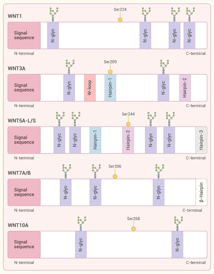
Figure 1.
Structural features specific to representative Wnt proteins are outlined
-
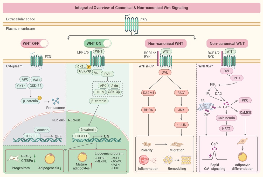
Figure 2.
Canonical and non-canonical Wnt signaling pathways
-
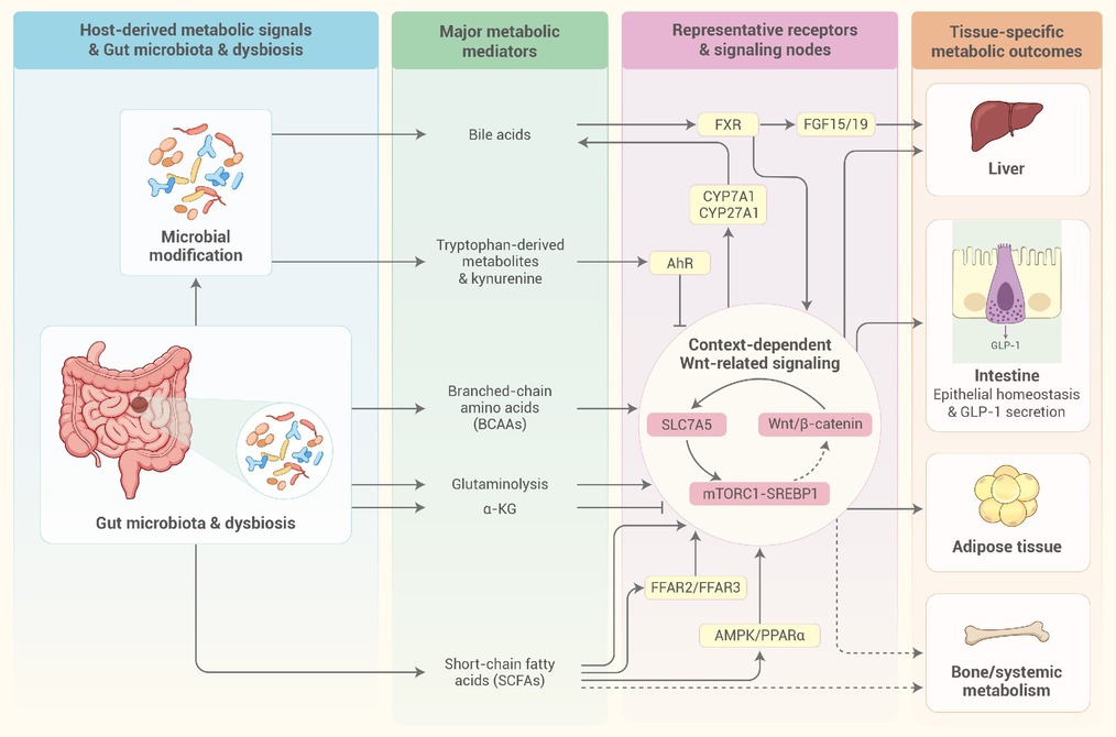
Figure 3.
Context-dependent interactions among metabolites derived from gut microbes, Wnt-related signaling, and lipid metabolism
-
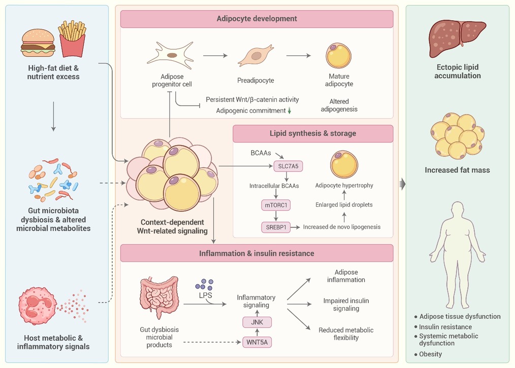
Figure 4.
Wnt-related mechanisms contributing to obesity and adipose tissue dysfunction
