
-
GRAPHICAL ABSTRACT
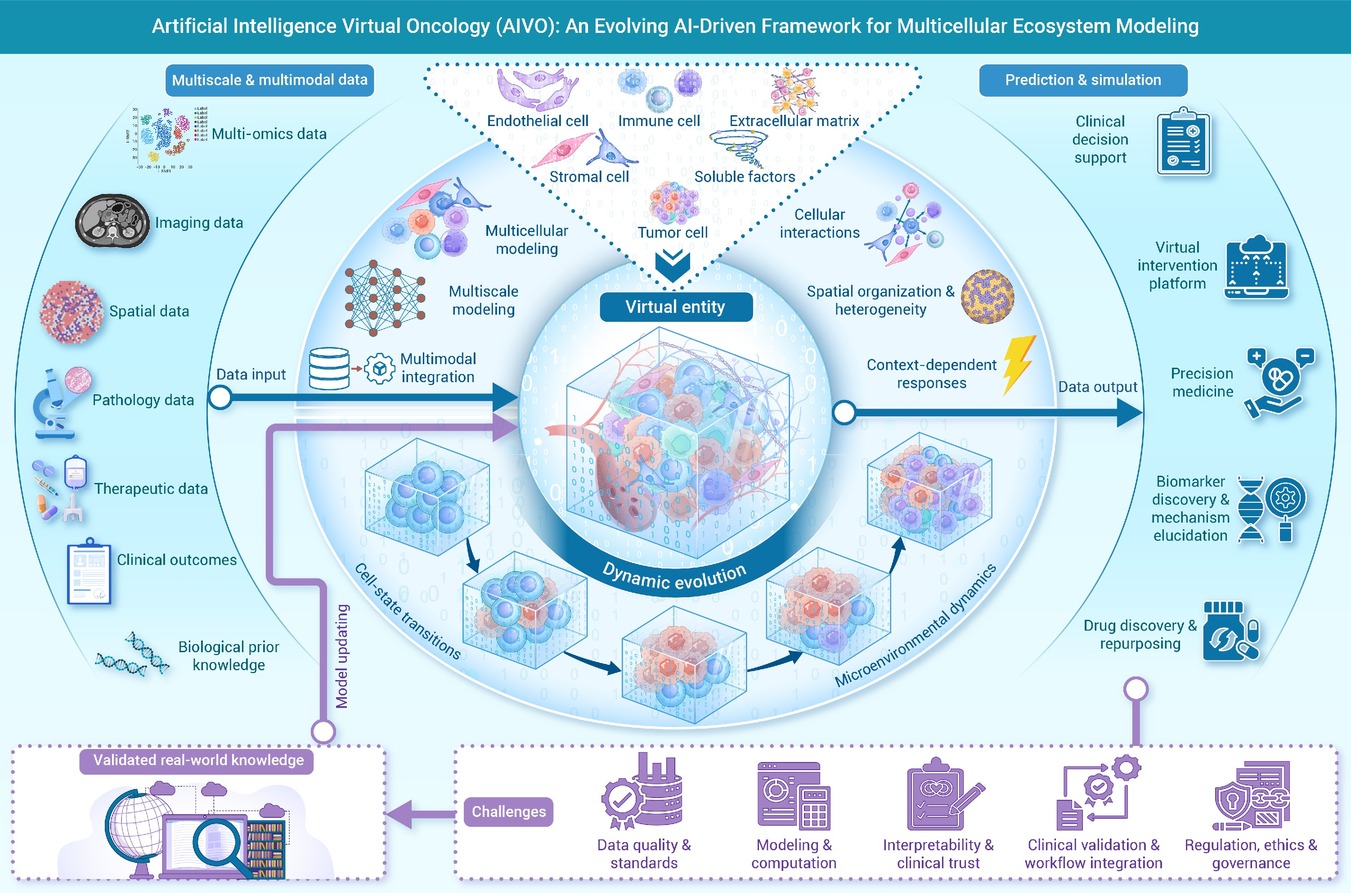
 DownLoad:
Full size image
DownLoad:
Full size image
-
PUBLIC SUMMARY
-
Artificial intelligence virtual oncology (AIVO) extends individual-cell modeling to tumor ecosystems.
AIVO as a virtual entity uses dynamic simulation to improve diagnosis and treatment for precision oncology.
AIVO remains conceptual, facing challenges in standards, reproducibility, validation, regulation, and equity.
Further interdisciplinary research and real-world validation are needed before AIVO becomes a reliable tool.
-
-
ABSTRACT
Tumors are dynamic multicellular ecosystems in which heterogeneity, evolution, and cross-scale interactions remain major barriers to precision oncology. Although multiple technological advances have improved tumor characterization, they remain largely static and descriptive, limiting their ability to capture state transitions, treatment-induced remodeling, and evolving intercellular communication. Artificial intelligence virtual cell (AIVC) provides a foundation for modeling cellular states, perturbation responses, and functional outputs from multimodal data. We extend this concept to artificial intelligence virtual oncology (AIVO), a framework designed to reconstruct multicellular communication, coordinated evolution, and system-level remodeling among tumor, immune, and stromal compartments. This review summarizes the conceptual basis, construction strategies, and emerging applications of this progression from AIVC to AIVO in tumor diagnosis, dynamic monitoring, treatment response prediction, and drug screening. We further discuss key challenges in data integration, interpretability, patient-specific calibration, and clinical validation. Grounded in AIVC-based modeling, we propose AIVO as an emerging framework for multimodal integration, dynamic modeling, and decision support in future oncology.-
Keywords:
- Oncology/ /
- Multicellular /
- communication/ /
- Artificial /
- intelligence/ /
- Artificial /
- intelligence /
- virtual /
- cell/ /
- Artificial /
- intelligence /
- virtual /
- oncology
-

-
REFERENCES
[1] Brown J.S., Amend S.R., Austin R.H., et al. (2023). Updating the definition of cancer. Mol. Cancer Res. 21:1142−1147. DOI:10.1158/1541-7786.MCR-23-0411 [2] Macklin D.N., Ruggero N.A. and Covert M.W. (2014). The future of whole-cell modeling. Curr. Opin. Biotechnol. 28:111−115. DOI:10.1016/j.copbio.2014.01.012 [3] Karr J.R., Sanghvi J.C., Macklin D.N., et al. (2012). A whole-cell computational model predicts phenotype from genotype. Cell 150:389−401. DOI:10.1016/j.cell.2012.05.044 [4] Margulies M., Egholm M., Altman W.E., et al. (2005). Genome sequencing in microfabricated high-density picolitre reactors. Nature 437:376−380. DOI:10.1038/nature03959 [5] Satam H., Joshi K., Mangrolia U., et al. (2023). Next-generation sequencing technology: Current trends and advancements. Biology (Basel) 12:997. DOI:10.3390/biology12070997 [6] Bai L. and Su J. (2025). Artificial intelligence virtual organoids (AIVOs). Bioact. Mater. 59:45−68. DOI:10.1016/j.bioactmat.2025.12.030 [7] Bunne C., Roohani Y., Rosen Y., et al. (2024). How to build the virtual cell with artificial intelligence: Priorities and opportunities. Cell 187:7045−7063. DOI:10.1016/j.cell.2024.11.015 [8] Jiang H., Huang X., Bi X., et al. (2026). Artificial intelligence-enabled multi-scale virtual cell: Perspective, challenges, and opportunities. Briefings Bioinf. 27:bbag104. DOI:10.1093/bib/bbag104 [9] Schaff J., Fink C.C., Slepchenko B., et al. (1997). A general computational framework for modeling cellular structure and function. Biophys. J. 73:1135−1146. DOI:10.1016/S0006-3495(97)78146-3 [10] Schaff J. and Loew L.M. (1999). The virtual cell. Pac. Symp. Biocomput. 228–239. DOI:10.1142/9789814447300_0023. [11] Tomita M., Hashimoto K., Takahashi K., et al. (1999). E-CELL: Software environment for whole-cell simulation. Bioinformatics 15:72−84. DOI:10.1093/bioinformatics/15.1.72 [12] Ahn-Horst T.A., Mille L.S., Sun G., et al. (2022). An expanded whole-cell model of E. coli links cellular physiology with mechanisms of growth rate control. NPJ Syst. Biol. Appl. 8:30. DOI:10.1038/s41540-022-00242-9 [13] Saliba A.E., Westermann A.J., Gorski S.A., et al. (2014). Single-cell RNA-seq: Advances and future challenges. Nucleic Acids Res. 42:8845−8860. DOI:10.1093/nar/gku555 [14] Boutros M., Heigwer F. and Laufer C. (2015). Microscopy-based high-content screening. Cell 163:1314−1325. DOI:10.1016/j.cell.2015.11.007 [15] Shalem O., Sanjana N.E., Hartenian E., et al. (2014). Genome-scale CRISPR-Cas9 knockout screening in human cells. Science 343:84−87. DOI:10.1126/science.1247005 [16] Dixit A., Parnas O., Li B., et al. (2016). Perturb-seq: Dissecting molecular circuits with scalable single-cell RNA profiling of pooled genetic screens. Cell 167:1853−1866. DOI:10.1016/j.cell.2016.11.038 [17] Rozenblatt-Rosen O., Stubbington M.J.T., Regev A., et al. (2017). The Human Cell Atlas: From vision to reality. Nature 550:451−453. DOI:10.1038/550451a [18] Lopez R., Regier J., Cole M.B., et al. (2018). Deep generative modeling for single-cell transcriptomics. Nat. Methods 15:1053−1058. DOI:10.1038/s41592-018-0229-2 [19] Jumper J., Evans R., Pritzel A., et al. (2021). Highly accurate protein structure prediction with AlphaFold. Nature 596:583−589. DOI:10.1038/s41586-021-03819-2 [20] Cui H., Wang C., Maan H., et al. (2024). scGPT: Toward building a foundation model for single-cell multi-omics using generative AI. Nat. Methods 21:1470−1480. DOI:10.1038/s41592-024-02201-0 [21] Heimberg G., Kuo T., DePianto D.J., et al. (2025). A cell atlas foundation model for scalable search of similar human cells. Nature 638:1085−1094. DOI:10.1038/s41586-024-08411-y [22] Hao M., Gong J., Zeng X., et al. (2024). Large-scale foundation model on single-cell transcriptomics. Nat. Methods 21:1481−1491. DOI:10.1038/s41592-024-02305-7 [23] Roohani Y.H., Hua T.J., Tung P.Y., et al. (2025). Virtual cell challenge: Toward a turing test for the virtual cell. Cell 188:3370−3374. DOI:10.1016/j.cell.2025.06.008 [24] Qian L., Dong Z. and Guo T. (2025). Grow AI virtual cells: Three data pillars and closed-loop learning. Cell Res. 35:319−321. DOI:10.1038/s41422-025-01101-y [25] Noutahi E., Hartford J., Tossou P., et al. (2025). Virtual cells: Predict, explain, discover. Preprint at arXiv. DOI:10.48550/arXiv.2505.14613. [26] Tomek J., Holmes M., Bury T., et al. (2026). T-World virtual human cardiomyocyte. I. development, validation, and cell arrhythmogenesis . Circ. Res. 138:e328073. DOI:10.1161/CIRCRESAHA.125.328073 [27] Thornburg Z.R., Maytin A., Kwon J., et al. (2026). Bringing the genetically minimal cell to life on a computer in 4D. Cell 189:2582−2597.e2527. DOI:10.1016/j.cell.2026.02.009 [28] Li W., Chen Y., Peng Z., et al. (2026). AetherCell: A generative engine for virtual cell perturbation and in vivo drug discovery. Preprint at bioRxiv. DOI:10.64898/2026.03.13.710968. [29] Chuai G., Chen X., Yang X., et al. (2026). Towards building a World Model to simulate perturbation-induced cellular dynamics by AlphaCell. Preprint at bioRxiv. DOI:10.64898/2026.03.02.709176. [30] Zhang H., Yuan G.H., Yuan C., et al. (2026). Lingshu-Cell: A generative cellular world model for transcriptome modeling toward virtual cells. Preprint at arXiv. DOI:10.48550/arXiv.2603.25240. [31] Chen S., Yu L., Jin K., et al. (2026). SCALE: Scalable conditional atlas-level endpoint transport for virtual cell perturbation prediction. Preprint at arXiv. DOI:10.48550/arXiv.2603.17380. [32] He S., Zhu Y., Tavakol D.N., et al. (2025). Squidiff: Predicting cellular development and responses to perturbations using a diffusion model. Nat. Methods 23:65−77. DOI:10.1038/s41592-025-02877-y [33] Zhang Y., Su Y., Wefers Z., et al. (2026). CellFluxV2: An image generative foundation model for virtual cell modeling. Preprint at bioRxiv. DOI:10.64898/2026.01.19.696785. [34] Wu D., Su S., Zhang Y., et al. (2026). CellFluxRL: Biologically-constrained virtual cell modeling via reinforcement learning. Preprint at arXiv. DOI:10.48550/arXiv.2603.21743. [35] Wang W., Hu Z., Weiler P., et al. (2026). RegVelo: Gene-regulatory-informed dynamics of single cells. Cell 189:3773−3800.e3744. DOI:10.1016/j.cell.2026.04.022 [36] Ellington C.N., Addagudi S., Wang J., et al. (2026). Cell-level virtual screening. Preprint at bioRxiv. DOI:10.64898/2026.05.11.724149. [37] Weidener L., Brkić M., Jovanović M., et al. (2026). VCBench: A multi-dimensional benchmark for single-cell foundation models. Preprint at bioRxiv. DOI:10.64898/2026.06.18.733146. [38] Rood J.E., Hupalowska A. and Regev A. (2024). Toward a foundation model of causal cell and tissue biology with a perturbation cell and tissue atlas. Cell 187:4520−4545. DOI:10.1016/j.cell.2024.07.035 [39] Ma C., Zhang H., Rao Y., et al. (2025). AI-driven virtual cell models in preclinical research: Technical pathways, validation mechanisms, and clinical translation potential. npj Digit. Med. 9:25. DOI:10.1038/s41746-025-02198-6 [40] Wei R., Wang B., Yan B., et al. (2026). From equations to agents: The artificial intelligence virtual cell reshaping precision oncology. Innov. Oncol. 1:100002. DOI:10.59717/j.xinn-oncol.2026.100002 [41] Yang T., Wang Y.Y., Ma F., et al. (2025). Build the virtual cell with artificial intelligence: A perspective for cancer research. Mil. Med. Res. 12:4. DOI:10.1186/s40779-025-00591-6 [42] Li M., Liu J., Xu Z., et al. (2026). Application prospects of artificial intelligence-driven virtual cell in reshaping the diagnostic and therapeutic paradigm of osteoarthritis. O.T.E. 1:025350013. DOI:10.36922/OTE025350013 [43] Baek S., Song K. and Lee I. (2025). Single-cell foundation models: Bringing artificial intelligence into cell biology. Exp. Mol. Med. 57:2169−2181. DOI:10.1038/s12276-025-01547-5 [44] Imianowski C.J., Chen Q., Workman C.J., et al. (2025). Regulatory T cells in the tumour microenvironment. Nat. Rev. Cancer 25:703−722. DOI:10.1038/s41568-025-00832-9 [45] Gavish A., Tyler M., Greenwald A.C., et al. (2023). Hallmarks of transcriptional intratumour heterogeneity across a thousand tumours. Nature 618:598−606. DOI:10.1038/s41586-023-06130-4 [46] Stevens A.J., Harris A.R., Gerdts J., et al. (2023). Programming multicellular assembly with synthetic cell adhesion molecules. Nature 614:144−152. DOI:10.1038/s41586-022-05622-z [47] Wegmann R., Bonilla X., Casanova R., et al. (2024). Single-cell landscape of innate and acquired drug resistance in acute myeloid leukemia. Nat. Commun. 15:9402. DOI:10.1038/s41467-024-53535-4 [48] Wang J., Wang T., Han R., et al. (2025). Artificial intelligence in cancer pathology: Applications, challenges, and future directions. Cytojournal 22:45. DOI:10.25259/Cytojournal_272_2024 [49] Shen S., Qi W., Liu X., et al. (2025). From virtual to reality: innovative practices of digital twins in tumor therapy. J. Transl. Med. 23:348. DOI:10.1186/s12967-025-06371-z [50] Ding J., Sharon N. and Bar-Joseph Z. (2022). Temporal modelling using single-cell transcriptomics. Nat. Rev. Genet. 23:355−368. DOI:10.1038/s41576-021-00444-7 [51] Xu H., Usuyama N., Bagga J., et al. (2024). A whole-slide foundation model for digital pathology from real-world data. Nature 630:181−188. DOI:10.1038/s41586-024-07441-w [52] Ding T., Wagner S.J., Song A.H., et al. (2025). A multimodal whole-slide foundation model for pathology. Nat. Med. 31:3749−3761. DOI:10.1038/s41591-025-03982-3 [53] Shulman E.D., Campagnolo E.M., Lodha R., et al. (2026). AI-predicted spatial transcriptomics unlocks breast cancer biomarkers from pathology. Cell 189:4225−4240. DOI:10.1016/j.cell.2026.04.023 [54] Pérez-Benito Á., García-Aznar J.M., Gómez-Benito M.J., et al. (2024). Patient-specific prostate tumour growth simulation: A first step towards the digital twin. Front. Physiol. 15:1421591. DOI:10.3389/fphys.2024.1421591 [55] Camacho-Gomez D., Borau C., Garcia-Aznar J.M., et al. (2025). Physics-informed machine learning digital twin for reconstructing prostate cancer tumor growth via PSA tests. npj Digit. Med. 8:485. DOI:10.1038/s41746-025-01890-x [56] Becker S., Mang A., Toma A., et al. (2010). In-silico oncology: An approximate model of brain tumor mass effect based on directly manipulated free form deformation. Int. J. Comput. Assist. Radiol. Surg. 5:607−622. DOI:10.1007/s11548-010-0531-7 [57] Stamatakos G.S. and Giatili S.G. (2017). A numerical handling of the boundary conditions imposed by the skull on an inhomogeneous diffusion-reaction model of glioblastoma invasion into the brain: Clinical validation aspects. Cancer Inform. 16:1176935116684824. DOI:10.1177/1176935116684824. [58] Antonopoulos M. and Stamatakos G. (2015). In silico neuro-oncology: Brownian motion-based mathematical treatment as a potential platform for modeling the infiltration of glioma cells into normal brain tissue. Cancer Inform. 14:33−40. DOI:10.4137/CIN.S19341 [59] Tomic H., Bjerkén A., Hellgren G., et al. (2022). Development and evaluation of a method for tumor growth simulation in virtual clinical trials of breast cancer screening. J. Med. Imaging 9:033503. DOI:10.1117/1.Jmi.9.3.033503 [60] Meyerheim M., Panagiotidou F., Georgiadi E., et al. (2025). Exploring the in silico adaptation of the nephroblastoma oncosimulator to MRI scans, treatment data, and histological profiles of patients from different risk groups. Front. Physiol. 16:1465631. DOI:10.3389/fphys.2025.1465631 [61] Stamatakos G.S., Antipas V.P. and Uzunoglu N.K. (2006). Simulating chemotherapeutic schemes in the individualized treatment context: The paradigm of glioblastoma multiforme treated by temozolomide in vivo. Comput. Biol. Med. 36:1216−1234. DOI:10.1016/j.compbiomed.2005.06.002 [62] Wu C., Jarrett A.M., Zhou Z., et al. (2022). MRI-based digital models forecast patient-specific treatment responses to neoadjuvant chemotherapy in triple-negative breast cancer. Cancer Res. 82:3394−3404. DOI:10.1158/0008-5472.CAN-22-1329 [63] Kolokotroni E., Dionysiou D., Veith C., et al. (2016). In silico oncology: Quantification of the in vivo antitumor efficacy of cisplatin-based doublet therapy in non-small cell lung cancer (NSCLC) through a multiscale mechanistic model. PLoS Comput. Biol. 12:e1005093. DOI:10.1371/journal.pcbi.1005093 [64] Demir S., Kessler T., Hotes A., et al. (2025). Mechanistic models position ceritinib as a nuclear integrity disrupting therapy in pediatric liver tumors. J. Exp. Clin. Cancer Res. 44:268. DOI:10.1186/s13046-025-03535-z [65] Gasselhuber A., Dreher M.R., Rattay F., et al. (2012). Comparison of conventional chemotherapy, stealth liposomes and temperature-sensitive liposomes in a mathematical model. PLoS One 7:e47453. DOI:10.1371/journal.pone.0047453 [66] Luu K.T. and Uchizono J.A. (2005). P-glycoprotein induction and tumor cell-kill dynamics in response to differential doxorubicin dosing strategies: A theoretical pharmacodynamic model. Pharm. Res. 22:710−715. DOI:10.1007/s11095-005-2585-8 [67] Liu C., Krishnan J. and Xu X.Y. (2013). Investigating the effects of ABC transporter-based acquired drug resistance mechanisms at the cellular and tissue scale. Integr. Biol. 5:555−568. DOI:10.1039/c2ib20238g [68] Derippe T., Fouliard S., Decleves X., et al. (2024). Quantitative systems pharmacology modeling of tumor heterogeneity in response to BH3‐mimetics using virtual tumors calibrated with cell viability assays. CPT: Pharmacometrics Syst. Pharmacol. 13:1252−1263. DOI:10.1002/psp4.13158 [69] Alshorman J., Mehran M.J., Bahrami Y., et al. (2026). Artificial intelligence in immunotherapy: Revolutionizing diagnostic and therapeutic applications in cancer and autoimmune diseases. Clin. Exp. Med. 26:185. DOI:10.1007/s10238-026-02107-5 [70] Gao D., Kan P., He Y., et al. (2025). Senescent immune cells in the tumor microenvironment: Emerging insights into cancer immunotherapy resistance. Front. Immunol. 16:1656733. DOI:10.3389/fimmu.2025.1656733 [71] Aung T.N., Monkman J., Warrell J., et al. (2025). Spatial signatures for predicting immunotherapy outcomes using multi-omics in non-small cell lung cancer. Nat. Genet. 57:2482−2493. DOI:10.1038/s41588-025-02351-7 [72] Zhai M., Zhang Z., Dong J., et al. (2025). Spatial proteomic profiling reveals conserved prognostic immune microenvironment features across molecular subtypes in small cell lung cancer. Pharmacol. Res. 222:108048. DOI:10.1016/j.phrs.2025.108048 [73] Stein-O'Brien G.L., Le D.T., Jaffee E.M., et al. (2023). Converging on a cure: The roads to predictive immunotherapy. Cancer Discovery 13:1053−1057. DOI:10.1158/2159-8290.CD-23-0277 [74] Wang Y., Bergman D.R., Trujillo E., et al. (2024). Agent-based modeling of virtual tumors reveals the critical influence of microenvironmental complexity on immunotherapy efficacy. Cancers 16:2942. DOI:10.3390/cancers16172942 [75] Gong C., Milberg O., Wang B., et al. (2017). A computational multiscale agent-based model for simulating spatio-temporal tumour immune response to PD1 and PDL1 inhibition. J. R. Soc. Interface 14:20170320. DOI:10.1098/rsif.2017.0320 [76] Kather J.N., Poleszczuk J., Suarez-Carmona M., et al. (2017). In silico modeling of immunotherapy and stroma-targeting therapies in human colorectal cancer. Cancer Res. 77:6442−6452. DOI:10.1158/0008-5472.CAN-17-2006 [77] Mösch A., Grazioli F., Machart P., et al. (2024). NeoAgDT: Optimization of personal neoantigen vaccine composition by digital twin simulation of a cancer cell population. Bioinformatics 40:btae205. DOI:10.1093/bioinformatics/btae205 [78] Bosque J.J., Martínez J., Otero J.G., et al. (2025). Mathematical model of tumor-macrophage dynamics in glioma to advance myeloid-targeted therapies. Comput. Biol. Med. 196:110909. DOI:10.1016/j.compbiomed.2025.110909 [79] Susilo M.E., Li C.C., Gadkar K., et al. (2023). Systems-based digital twins to help characterize clinical dose-response and propose predictive biomarkers in a phase I study of bispecific antibody, mosunetuzumab, in NHL. Clin. Transl. Sci. 16:1134−1148. DOI:10.1111/cts.13501 [80] Wang Y., Casarin S., Daher M., et al. (2026). Agent-based modeling of cellular dynamics in adoptive cell therapy. Commun. Biol. 9:409. DOI:10.1038/s42003-026-09653-4 [81] Pan X., Li X., Dong L., et al. (2024). Tumour vasculature at single-cell resolution. Nature 632:429−436. DOI:10.1038/s41586-024-07698-1 [82] Wang X., Li S., Xue Y., et al. (2026). The heterogeneity and function of stromal cells in the tumor microenvironment. Research 9:1183. DOI:10.34133/research.1183 [83] Cho M., Koh E.Y., Kim Y., et al. (2025). Molecular profiling of inflammatory and myofibroblast cancer-associated fibroblast subtypes derived from human pancreatic stellate cells using machine learning-based label-free raman spectroscopy. Biomater. Res. 29:0292. DOI:10.34133/bmr.0292 [84] Laursen R., Chen H., Demaray J., et al. (2026). Neighborhood nonnegative matrix factorization identifies patterns and spatially-variable genes in large-scale spatial transcriptomics data. Genome Biol. 27:31. DOI:10.1186/s13059-025-03846-6 [85] Ma A., Wang X., Li J., et al. (2023). Single-cell biological network inference using a heterogeneous graph transformer. Nat. Commun. 14:964. DOI:10.1038/s41467-023-36559-0 [86] Johnson J.A.I., Bergman D.R., Rocha H.L., et al. (2025). Human interpretable grammar encodes multicellular systems biology models to democratize virtual cell laboratories. Cell 188:4711−4733.e4737. DOI:10.1016/j.cell.2025.06.048 [87] Lv Y., Duan T., Song J., et al. (2026). The spatiotemporal heterogeneity of tumor-associated stromal cells: Reprogramming plasticity to unlock precision cancer immunotherapy. Cancer Commun. 46:0002. DOI:10.34133/cancomm.0002 [88] Lee J. and Kim E. (2025). Ordinary differential equation model of cancer-associated fibroblast heterogeneity predicts treatment outcomes. npj Syst. Biol. Appl. 11:96. DOI:10.1038/s41540-025-00578-y [89] Zhang S., Wang H., Cho Y., et al. (2025). Quantitative calibration of a spatial QSP model identifies fibroblast impact on HCC immunotherapy. Preprint at bioRxiv. DOI:10.1101/2025.10.31.685939. [90] Kalluri R. (2016). The biology and function of fibroblasts in cancer. Nat. Rev. Cancer 16:582−598. DOI:10.1038/nrc.2016.73 [91] Zhang F., Ma Y., Li D., et al. (2024). Cancer associated fibroblasts and metabolic reprogramming: Unraveling the intricate crosstalk in tumor evolution. J Hematol. Oncol. 17:80. DOI:10.1186/s13045-024-01600-2 [92] McAllister S.S. and Weinberg R.A. (2010). Tumor-host interactions: A far-reaching relationship. J. Clin. Oncol. 28:4022−4028. DOI:10.1200/JCO.2010.28.4257 [93] Metzcar J., Duggan B.S., Fischer B., et al. (2025). A simple framework for agent-based modeling with extracellular matrix. Bull. Math. Biol. 87:43. DOI:10.1007/s11538-024-01408-8 [94] Biancalani T., Scalia G., Buffoni L., et al. (2021). Deep learning and alignment of spatially resolved single-cell transcriptomes with Tangram. Nat. Methods 18:1352−1362. DOI:10.1038/s41592-021-01264-7 [95] Kleshchevnikov V., Shmatko A., Dann E., et al. (2022). Cell2location maps fine-grained cell types in spatial transcriptomics. Nat. Biotechnol. 40:661−671. DOI:10.1038/s41587-021-01139-4 [96] Cable D.M., Murray E., Zou L.S., et al. (2022). Robust decomposition of cell type mixtures in spatial transcriptomics. Nat. Biotechnol. 40:517−526. DOI:10.1038/s41587-021-00830-w [97] Hu J., Li X., Coleman K., et al. (2021). SpaGCN: Integrating gene expression, spatial location and histology to identify spatial domains and spatially variable genes by graph convolutional network. Nat. Methods 18:1342−1351. DOI:10.1038/s41592-021-01255-8 [98] Dong K. and Zhang S. (2022). Deciphering spatial domains from spatially resolved transcriptomics with an adaptive graph attention auto-encoder. Nat. Commun. 13:1739. DOI:10.1038/s41467-022-29439-6 [99] Jin S., Guerrero-Juarez C.F., Zhang L., et al. (2021). Inference and analysis of cell-cell communication using CellChat. Nat. Commun. 12:1088. DOI:10.1038/s41467-021-21246-9 [100] Browaeys R., Saelens W. and Saeys Y. (2020). NicheNet: Modeling intercellular communication by linking ligands to target genes. Nat. Methods 17:159−162. DOI:10.1038/s41592-019-0667-5 [101] Cang Z., Zhao Y., Almet A.A., et al. (2023). Screening cell-cell communication in spatial transcriptomics via collective optimal transport. Nat. Methods 20:218−228. DOI:10.1038/s41592-022-01728-4 [102] Yang Z., Su W., Wei X., et al. (2025). Hypoxia inducible factor-1α drives cancer resistance to cuproptosis. Cancer Cell 43:937−954. DOI:10.1016/j.ccell.2025.02.015 [103] Liu L., Zhang Y., Liu H., et al. (2025). Transforming cancer therapy: Unlocking the potential of targeting vascular and stromal cells in the tumor microenvironment. Cancer Res. 85:2152−2164. DOI:10.1158/0008-5472.CAN-24-4744 [104] Zhang S.W., Wang H., Xiao Y., et al. (2026). Sensory neurons drive immune exclusion by stimulating a dense extracellular matrix in the breast cancer tumor microenvironment. Cell 189:1039−1055. DOI:10.1016/j.cell.2026.01.001 [105] Hunter M.V., Joshi E., Bowker S., et al. (2025). Mechanical confinement governs phenotypic plasticity in melanoma. Nature 647:517−527. DOI:10.1038/s41586-025-09445-6 [106] Semenza G.L. (2002). HIF-1 and tumor progression: Pathophysiology and therapeutics. Trends. Mol. Med. 8:S62−S67. DOI:10.1016/s1471-4914(02)02317-1 [107] Yuan X., Ruan W., Bobrow B., et al. (2024). Targeting hypoxia-inducible factors: Therapeutic opportunities and challenges. Nat. Rev. Drug. Discov. 23:175−200. DOI:10.1038/s41573-023-00848-6 [108] Schiavo F., Lindblom E.K. and Toma-Dasu I. (2022). Towards the virtual tumor for optimizing radiotherapy treatments of hypoxic tumors: A novel model of heterogeneous tissue vasculature and oxygenation. J. Theor. Biol. 547:111175. DOI:10.1016/j.jtbi.2022.111175 [109] Nissen N.I., Karsdal M. and Willumsen N. (2019). Collagens and cancer associated fibroblasts in the reactive stroma and its relation to cancer biology. J. Exp. Clin. Cancer Res. 38:115. DOI:10.1186/s13046-019-1110-6 [110] Swat M.H., Thomas G.L., Shirinifard A., et al. (2015). Emergent stratification in solid tumors selects for reduced cohesion of tumor cells: A multi-cell, virtual-tissue model of tumor evolution using CompuCell3D. PLoS One 10:e0127972. DOI:10.1371/journal.pone.0127972 [111] Hendriks B.S., Reynolds J.G., Klinz S.G., et al. (2012). Multiscale kinetic modeling of liposomal doxorubicin delivery quantifies the role of tumor and drug‐specific parameters in local delivery to tumors. CPT: Pharmacometrics Syst. Pharmacol. 1:e15. DOI:10.1038/psp.2012.16 [112] González-Garcinuño Á., Valle E.M.D. and Kenjeres S. (2026). From blood flow to tumor cell internalization: A multistage computational model of nanoparticle dynamics. Pharm. Res. 43:405−419. DOI:10.1007/s11095-026-04018-2 [113] Aghamiri S.S. and Amin R. (2025). The potential use of digital twin technology for advancing CAR-T cell therapy. Curr. Issues Mol. Biol. 47:321. DOI:10.3390/cimb47050321 [114] Karaman I. and Sebin B. (2025). From data-driven cities to data-driven tumors: Dynamic digital twins for adaptive oncology. Front. Artif. Intell. 8:1624877. DOI:10.3389/frai.2025.1624877 [115] Nardone V., Marmorino F., Germani M.M., et al. (2024). The Role of artificial intelligence on tumor boards: Perspectives from surgeons, medical oncologists and radiation oncologists. Curr. Oncol. 31:4984−5007. DOI:10.3390/curroncol31090369 [116] Malerba S., Vladimirov M., Goyal A., et al. (2026). Artificial intelligence applications in gastric cancer surgery: Bridging early diagnosis and responsible precision medicine. J. Clin. Med. 15:2208. DOI:10.3390/jcm15062208 [117] Goyal A., Koutentakis M., Park J., et al. (2025). Artificial intelligence for risk-benefit assessment in hepatopancreatobiliary oncologic surgery: A systematic review of current applications and future directions on behalf of TROGSS-The Robotic Global Surgical Society. Cancers (Basel) 17:3292. DOI:10.3390/cancers17203292 [118] Ardila C.M. and Yadalam P.K. (2025). Unresolved questions in the application of artificial intelligence virtual cells for cancer research. Mil. Med. Res. 12:19. DOI:10.1186/s40779-025-00608-0 [119] Li J., Zhang H., Zhang W., et al. (2026). Virtual cell: Current perspectives and future prospects. J. Int. Med. Res. 54:3000605261425080. DOI:10.1177/03000605261425080. [120] Chua I.S., Gaziel-Yablowitz M., Korach Z.T., et al. (2021). Artificial intelligence in oncology: Path to implementation. Cancer Med. 10:4138−4149. DOI:10.1002/cam4.3935 [121] Lotter W., Hassett M.J., Schultz N., et al. (2024). Artificial intelligence in oncology: Current landscape, challenges, and future directions. Cancer Discov. 14:711−726. DOI:10.1158/2159-8290.CD-23-1199 [122] Lin R., Zhao Z., Liu Z., et al. (2026). Artificial intelligence in clinical oncology: Multimodal integration and translational development. Cancer Lett. 649:218493. DOI:10.1016/j.canlet.2026.218493 -
ABOUT THIS ARTICLE
Cite this article:
Lyu C., Yao D., Yin F., et al. (2026). Artificial intelligence derived virtual oncology. The Innovation Oncology 1:100028. https://doi.org/10.59717/j.xinn-oncol.2026.100028
Welcome!
To request copyright permission to republish or share portions of our works, please visit Copyright Clearance Center's (CCC) Marketplace website at marketplace.copyright.com.
Export File
Citation
Lyu C., Yao D., Yin F., et al. (2026). Artificial intelligence derived virtual oncology. The Innovation Oncology 1:100028. https://doi.org/10.59717/j.xinn-oncol.2026.100028

Format
Content
-
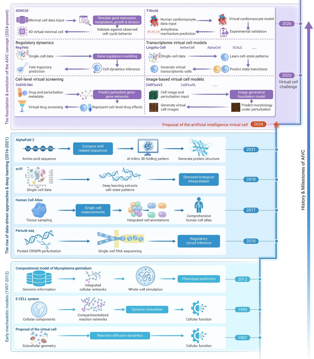
Figure 1.
History and milestones of AIVC
-
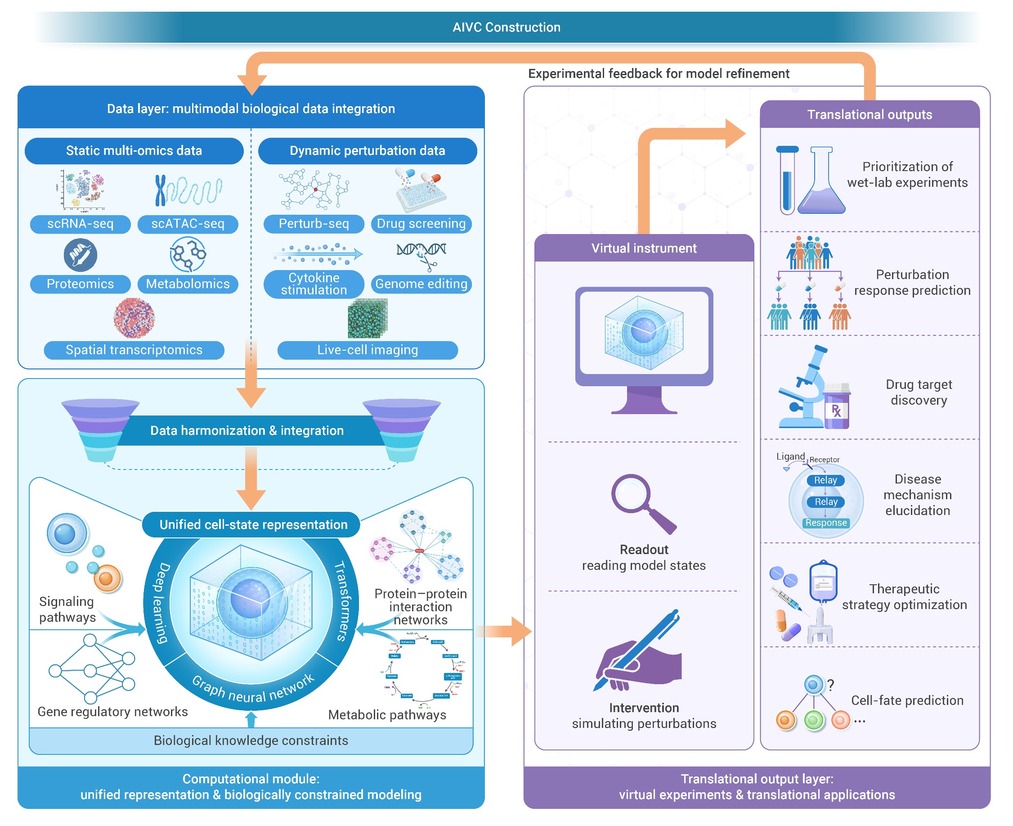
Figure 2.
The construction of AIVC
-
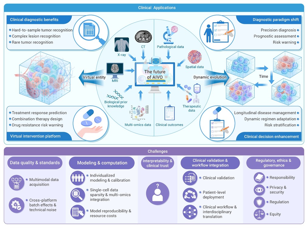
Figure 3.
Future directions and challenges of AIVO
